Support Chiropractic Research!

Orthopedic Tests

Sports Management: Shoulder Girdle Injuries

By |April 20, 2013|Chiropractic Care, Education, Orthopedic Tests, Rehabilitation, Shoulder Girdle Injuries, Sports Management|

Sports Management: Shoulder Girdle Injuries

The Chiro.Org Blog


We would all like to thank Dr. Richard C. Schafer, DC, PhD, FICC for his lifetime commitment to the profession. In the future we will continue to add materials from RC’s copyrighted books for your use.

This is Chapter 22 from RC’s best-selling book:

“Chiropractic Management of Sports and Recreational Injuries”

Second Edition ~ Wiliams & Wilkins

These materials are provided as a service to our profession. There is no charge for individuals to copy and file these materials. However, they cannot be sold or used in any group or commercial venture without written permission from ACAPress.


Chapter 22:   Shoulder Girdle Injuries

This chapter concerns injuries of and about the scapula, clavicle, and shoulder. In sports, the shoulder girdle is a common site of minor injury and a not infrequent site of serious disability. It is second only to the knee as a chronic site of prolonged disability. Upper limb injuries amount to about 20% of sport-related injuries. They can be highly debilitating, require considerable lost field time, and can easily ruin a promising sports career.


Introduction


The versatile shoulder girdle consists of the sternoclavicular, acromioclavicular, and glenohumeral joints, and the scapulothoracic articulation. These allow, as a whole, universal mobility by way of a shallow glenoid fossa, the joint capsule, and the suspension muscles and ligaments. The shoulder, a ball-and-socket joint, is freely movable and lacks a close connection between its articular surfaces.

The regional anatomy offers little to resist violent shoulder depression, and the shoulder tip itself has little protection from trauma. The length of the arm presents a long lever with a large head within a relatively small joint. This allows a great range of motion with little stability. The stability of the shoulder is derived entirely from its surrounding soft tissues.

History and Initial Care

A careful history recording the mechanism of trauma and the position of the limb during injury, careful inspection and palpation of the entire region, muscle and range-of-motion tests, and other standard neurologic-orthopedic tests will often arrive at an accurate diagnosis without the necessity of x-ray exposure. Forceful manipulations should always be reserved for late in the examination to evaluate contraindications.

Contusions, strains, sprains, bursitis, and neurologic deficits must be alertly recognized and treated. Fractures and dislocations, obviously, take precedence over soft-tissue injuries with the exception of severe bleeding. Always check for bony crepitus, fracture line tenderness and swelling, angulation and deformity. Because the shoulder readily “freezes” after injury, treatment must strive to maintain motion as soon as possible without encouraging recurring problems. The key to avoiding prolonged disability is early recognition and early mobilization.

There are more materials like this @ our:

Shoulder Girdle Page

Posttraumatic Assessment (more…)

Clinical Disorders and the Sensory System

By |April 11, 2013|Chiropractic Education, Diagnosis, Education, Evaluation & Management, General Health, Health Promotion, Neurology, Orthopedic Tests, Radiculopathy, Spinal Manipulation|

Clinical Disorders and the Sensory System

The Chiro.Org Blog


We would all like to thank Dr. Richard C. Schafer, DC, PhD, FICC for his lifetime commitment to the profession. In the future we will continue to add materials from RC’s copyrighted books for your use.

This is Chapter 4 from RC’s best-selling book:

“Basic Principles of Chiropractic Neuroscience”

These materials are provided as a service to our profession. There is no charge for individuals to copy and file these materials. However, they cannot be sold or used in any group or commercial venture without written permission from ACAPress.


Chapter 8: Clinical Disorders and the Sensory System

This chapter describes those sensory mechanisms, joint signals, and abnormal sensations (eg, pain, thermal abnormalities) that have particular significance within clinical diagnosis. The basis and differentiation of pain are described, as are the related subjects of trigger points and paresthesia. The chapter concludes with a description of the neurologic basis for the evaluation of the sensory system and the sensory fibers of the cranial nerves.


THE ANALYSIS OF PAIN
IN THE CLINICAL SETTING


Although all pain does not have organic causes, there is no such thing as “imagined” pain. Pain that can be purely isolated as a structural, functional, or an emotional effect is rare. More likely, all three are superimposed upon and interlaced with each other in various degrees of status. This is also true for neural, vascular, lymphatic, and hormonal mechanisms.

Common Causes of Pain and Paresthesia

The common causes of pain and paresthesia are:

(1) obvious direct trauma or injury;

(2) reflex origins in musculoskeletal lesions, which deep pressure often exaggerates, such as trigger areas;

(3) peripheral nerve injury (eg, causalgia), which results in an intense burning superficial pain;

(4) the presence of nerve inflammations and degeneration of the peripheral or CNS, which frequently cause other changes indicative of such lesions; (more…)

Orthopedic and Neurologic Procedures in Chiropractic

By |November 15, 2012|Chiropractic Care, Diagnosis, Neurology, Orthopedic Tests, Rehabilitation|

Orthopedic and Neurologic Procedures in Chiropractic

The Chiro.Org Blog


We would all like to thank Dr. Richard C. Schafer, DC, PhD, FICC for his lifetime commitment to the profession. In the future we will continue to add materials from RC’s copyrighted books for your use.

This is Chapter 3 from RC’s best-selling book:
“Basic Chiropractic Procedural Manual”

These materials are provided as a service to our profession. There is no charge for individuals to copy and file these materials. However, they cannot be sold or used in any group or commercial venture without written permission from ACAPress.


Chapter 3: Orthopedic and Neurologic Procedures in Chiropractic

This chapter presents the general diagnostic methods currently used in differential diagnosis of selected orthopedic and neurologic conditions.


SELECTED NEUROLOGIC PROBLEMS


Overview

The typical patient presents the challenge of differential diagnosis of a number of neurologic conditions. These range from a variety of peripheral neuritides that may be completely reversible to serious degenerations of the central nervous system.

The tendency of the geriatric patient to develop neurologic problems is often related to the aging process: loss of tissue elasticity, particularly that of the musculoskeletal system. This is manifested by greater rigidity of the spinal column with the appearance of fixation subluxations. These, together with dehydration and subsequent thinning of the intervertebral discs, predispose to radiculitis, neuritis, and vasomotor disturbances and metabolic effects on the cord and brain. The neurologic disturbances can be superimposed on already degenerating arteriosclerotic vessels and alter metabolism of the gastrointestinal and other systems, which may cause serious problems unless recognized early and prompt corrective measures are administered.

Types of Neuritides

Peripheral Neuritis

Peripheral neuritis is a general peripheral neuritis such as that which may be present in such disorders as diabetes, anemia, and vitamin deficiency. Diminution of all sensation will be noted, with proprioception affected most. A stocking distribution with an ill-defined border is commonly witnessed. Glove distribution may appear later, along with paresthesias in the distal areas of sensory distribution. The clinical picture does not conform to either dermatome or nerve patterns of distribution. The cause for this is unknown.

Local Neuritis (more…)

Lower Back Trauma (Lumbar Spine and Pelvis)

By |May 20, 2012|Chiropractic Care, Chronic Pain, Evidence-based Medicine, Low Back Pain, Orthopedic Tests, Rehabilitation, Spinal Manipulation|

Lower Back Trauma (Lumbar Spine and Pelvis)

The Chiro.Org Blog


Clinical Monograph 24

By R. C. Schafer, DC, PhD, FICC


Although it may be easier to teach anatomy by dividing the body into arbitrary parts, a misinterpretation can be created. For instance, we find clinically that the lumbar spine, sacrum, ilia, pubic bones, and hips work as a functional unit. Any disorder of one part immediately affects the function of the other parts. We should also keep in mind that an axial kinematic chain of weight-supporting segments extends from the occipital base to the soles of the feet.

Because the number of professional papers concerning the cause and diagnosis of low-back pain is voluminous, emphasis herein is placed on points that the author believes are important but not often emphasized in popular literature.


BACKGROUND


A wide assortment of muscle, tendon, ligament, bone, nerve, and vascular injuries in this area is witnessed during posttrauma care. As with other areas of the body, the first step in the posttrauma examination process is knowing the mechanism of injury if possible. Evaluation can be rapid and accurate with this knowledge.

Low-back disability rapidly demotivates productivity and athletic participation. The mechanism of injury is usually intrinsic rather than extrinsic. The cause can often be through overbending, a heavy steady lift, or a sudden release –all which primarily involve the muscles. IVD disorders are more often, but not exclusively, attributed to extrinsic blows and intrinsic wrenches. An accurate and complete history is invariably necessary to offer the best management and counsel.

Initial Assessment

A player injured on the field or a worker injured in the shop should never be moved until emergency assessment is completed. Once severe injury has been eliminated, transfer to a backboard can be made and further evaluation conducted at an aid station.

Neurologic Levels

Neurologic assessment should be made as soon as logical. Muscle tonus (flaccidity, rigidity, spasticity) by passive movements is determined. Voluntary power of each suspected group of muscles against resistance is tested, and the force is compared bilaterally. Check pupil size, ability to follow finger motion, and reaction to light. Cremasteric (L1–L2), patellar (L2–L4), gluteal (L4–S1), suprapatellar, Achilles (L5–S2), plantar (S1–S2), and anal (S5–Cx1) reflexes are evaluated. Patellar and ankle clonuses are noted. Coordination and sensation by gait, heel-to-knee and foot-to-buttock tests, and Romberg’s station test are checked. These are typical minimal evaluations.

Initial Assessment

Tenderness.   Tenderness is frequently found at the apices of spinal curves and not infrequently where one curve merges with another. Tenderness about spinous or transverse processes is usually of low intensity and suggests articular stress. Tenderness noted at the points of nerve exit from the spine and continuing in the pathway of the peripheral division of the nerves is a valuable aid in spinal analysis pointing to a foraminal lesion. However, the lack of tenderness is not a clear indication of lack of spinal dysfunction. Tenderness is a subjective symptom influenced by many individual structural, functional, and psychologic factors that can make it an unreliable sign. An area for clues sometimes overlooked is the presence and symmetry of lower-extremity pulses.

Keep in mind that lumbopelvic tenderness as well as pain can be referred from pelvic and lower abdominal viscera.

LUMBAR SUBLUXATION SYNDROMES

Functional revolts associated with subluxation syndromes can manifest as abnormalities in sensory interpretations and/or motor activities. These disturbances may be through one of two primary mechanisms: direct nerve disorders or be of a reflex nature.

Nerve Root Insults


Read the rest of this Full Text article now!


Enjoy the rest of Dr. Schafer’s Monographs at:

Rehabilitation Monograph Page

Stabilizing The Pelvis, Using the Modified Kemps and Straight Leg Raise Tests And Post-isometric Relaxation (PIR)

By |January 7, 2012|Modified Kemp's test, Orthopedic Tests, Pelvic Stability, Post-isometric Relaxation, Straight Leg Raise|

Stabilizing The Pelvis, Using the Modified Kemps and Straight Leg Raise Tests And Post-isometric Relaxation (PIR)

The Chiro.Org Blog


SOURCE:   Chiro.Org’s Low Back Pain Page


Dr. Leonard Faye reminds us in Chapter 6 of Schafer’s text “Motion Palpation” that:

“In all low-back pain cases, it is essential to test for
hamstring, quadriceps, and psoas length.”

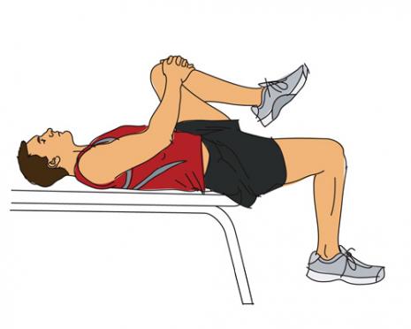
The picture on the left displays the positioning for the Modified Thomas Test. This test is is a very effective way to assess the length of 2 different muscles.

When the patient holds their knee to their chest, the following 2 things should become immediately apparent:

  1. If the psoas is of normal length, then the dependent thigh should be free to hang down 45° below the plane of the table. In this picture, the patient’s right psoas is significantly shortened.
  2. If the quadriceps are of normal length, the angle of the knee (between thigh and leg) should approximate 90°

If the psoas is shortened, it pulls the thigh into (some degree of) flexion, so the thigh cannot fully extend. This shortens your gait. Recalling that the origin of the psoas includes the lower thoracic vertebra, the lumbar segments I-IV, and the neighboring intervertebral discs, you can see why shortness would destabilize the lumbar and pelvic joints. Also… if the psoas is in contraction, the gluts may become inhibited by reciprocal inhibition.

If the quadriceps are shortened, it draws the leg into extension. Because portions of the quads originate on the pelvis, a shortened quads also distorts normal pelvic motion. (more…)