Support Chiropractic Research!

Education

Specific Potentialities of the Subluxation Complex

By |May 27, 2013|Cervical Spine, Diagnosis, Education, Subluxation|

Specific Potentialities of the Subluxation Complex

The Chiro.Org Blog


We would all like to thank Dr. Richard C. Schafer, DC, PhD, FICC for his lifetime commitment to the profession. In the future we will continue to add materials from RC’s copyrighted books for your use.

This is Chapter 7 from RC’s best-selling book:

“Basic Principles of Chiropractic Neuroscience”

These materials are provided as a service to our profession. There is no charge for individuals to copy and file these materials. However, they cannot be sold or used in any group or commercial venture without written permission from ACAPress.


Chapter 7: Specific Potentialities of the Subluxation Complex

This chapter describes the primary neurologic implications of subluxation syndromes, either as a primary factor or secondary to trauma or pathology, within the cervical spine, thoracic spine, lumbar spine, and pelvic articulations.


GENERAL CONSIDERATIONS


Studies reported by Drum, Hargrave-Wilson, Kunert, Burke, Gayral/Neuwirth, and others have shown that a subluxation complex, often leading to spondylosis, can effect a wide variety of disturbances that may appear to be disrelated on the surface. Most of the remote effects can be grouped under the general classifications of nerve root neuropathy, basilar venous congestion, cervical autonomic disturbances, CSF pressure and flow disturbances, axoplasmic flow blocks, irritation of the recurrent meningeal nerve, the Barre-Lieou syndrome, and/or the vertebral artery syndrome.

This chapter describes many causes for and effects of a spinal subluxation complex. In clinical practice, however, causes and effects are rarely found as isolated entities. Several factors will usually be involved and superimposed on each other.

Innervation of the Spinal Dura

It has long been known that the spinal dura mater has an intrinsic nerve supply. Spinal meningeal rami are derived from gray communicating rami and spinal nerves. The spinal nerves contribute sensory fibers to the meningeal rami. Several meningeal rami enter each IVF, and most are located anteriorly to the sensory ganglia within the IVF.

Bridge found that these intrinsic nerve fibers reach the anterior surface of the dura by three main courses. Here the nerves divide into ascending and usually longer descending filaments that run longitudinally and parallel on the dural surface, and a considerable amount of nerve overlaps from adjacent segments. Finer filaments penetrate the dural substance where they subdivide.

Kimmel reported that most of these fibers penetrate the dura near the midline, while others enter laterally near the exiting spinal nerve roots. At each segment level, two or three nerves enter the spinal dura mater and contain only small nerve fibers. In contrast, Edgar/Nundy could determine no definitive nerve endings, but the nerves could be traced to the posterior aspect of the spinal dura. These observations help to clarify the wide distribution of back pain that is often found following protrusion of a single IVD.

Cervical Dura Attachments

Sunderland states that the nerve sheaths in the cervical region are not firmly attached to their respective foramina. Only the C4 C6 cervical nerves have a strong attachment to the vertebral column, and this is to the gutter of the vertebral transverse process. He believes that these observations have relevance to any local lesion that may fix, deform, or otherwise affect the nerve and its nroots to the point of interfering with their function, and they also may be important to traction ijuries of nerve roots.
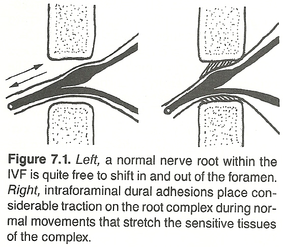
(more…)

Sports Management: Leg, Ankle, and Foot Injuries

By |April 23, 2013|Chiropractic Care, Chiropractic Education, Chiropractic Technique, Clinical Decision-making, Education, Gait Analysis, Rehabilitation, Sports Management|

Sports Management: Leg, Ankle, and Foot Injuries

The Chiro.Org Blog


We would all like to thank Dr. Richard C. Schafer, DC, PhD, FICC for his lifetime commitment to the profession. In the future we will continue to add materials from RC’s copyrighted books for your use.

This is Chapter 27 from RC’s best-selling book:

“Chiropractic Management of Sports and Recreational Injuries”

Second Edition ~ Wiliams & Wilkins

These materials are provided as a service to our profession. There is no charge for individuals to copy and file these materials. However, they cannot be sold or used in any group or commercial venture without written permission from ACAPress.


Chapter 27:   Leg, Ankle, and Foot Injuries

The lower leg, ankle, and foot work as a functional unit. Total body weight above is transmitted to the leg, ankle hinge, and foot in the upright position, and this force is greatly multiplied in locomotion. Thus the ankle and foot are uniquely affected by trauma and static deformities infrequently seen in other areas of the body.


Injuries of the Leg


The most common injuries in this area are bruises, muscle strains, tendon lesions, postural stress, anterior and posterior compression syndromes, and tibia and fibula fractures. Bruises of the lower leg are less frequent than those of the thigh or knee, but the incidence of intrinsic strain, sprain, and stress fractures are much greater.

A continual program of running and jogging is typical of most sports. The result is often strengthening of the antigravity muscles at the expense of the gravity muscles — producing a dynamic imbalance unless both gravity and antigravity muscles are developed simultaneously. An anatomic or physiologic short leg as little as an eighth of an inch can affect a stride and produce an overstrain in long-distance track events.

Bruises and Contusions

The most common bruise of the lower extremity is that of the shin where disability may be great as the poorly protected tibial periosteum is usually involved. Skin splits in this area can be most difficult to heal. Signs of suppuration indicate referral to guard against periostitis and osteomyelitis.

Management.   Treat as any skin-bone bruise with cold packs and antibacterial procedures, and shield the area with padding during competitive activity. When long socks are worn, the incidence of shinbone injuries is reduced. An old but effective protective method in professional football that does not add weight is to place four or five sheets of slick magazine pages around the shin that are secured by a cotton sock which is covered by the conventional sock. A blow to the shin is reduced to about a third of its force as the paper slips laterally on impact.

GASTROCNEMIUS CONTUSION

This is a common and most debilitating injury in contact sports. It is characterized by severe calf tenderness, abnormal muscle firmness of the engorged muscle, and inability to raise the heel during weight bearing.

Management.   Treat with cold packs, compression, and elevation for 24 hr. Follow with mild heat and contrast baths. Massage is contraindicated as it might disturb muscle repair. The danger of ossification is less in the calf than in the thigh, but management must incorporate precautions against adhesions.

TRAUMATIC PHLEBITIS

Contusion to the greater saphenous vein may lead to rupture resulting in extensive swelling, ecchymosis, redness and other signs of local phlebitis. Tenderness will be found along the course of the vascular channel. During treatment, referral should be made upon the first signs of thrombosis.

Management.   Management is by rest, cold, compression, and elevation for at least 24 hr. Later, progressive ambulation, mild heat, and contrast baths should be utilized. Progressive exercises may begin in 4-6 days. When competitive activity is resumed, the area should be provided extra protection.

NERVE CONTUSIONS (more…)

Sports Management: Shoulder Girdle Injuries

By |April 20, 2013|Chiropractic Care, Education, Orthopedic Tests, Rehabilitation, Shoulder Girdle Injuries, Sports Management|

Sports Management: Shoulder Girdle Injuries

The Chiro.Org Blog


We would all like to thank Dr. Richard C. Schafer, DC, PhD, FICC for his lifetime commitment to the profession. In the future we will continue to add materials from RC’s copyrighted books for your use.

This is Chapter 22 from RC’s best-selling book:

“Chiropractic Management of Sports and Recreational Injuries”

Second Edition ~ Wiliams & Wilkins

These materials are provided as a service to our profession. There is no charge for individuals to copy and file these materials. However, they cannot be sold or used in any group or commercial venture without written permission from ACAPress.


Chapter 22:   Shoulder Girdle Injuries

This chapter concerns injuries of and about the scapula, clavicle, and shoulder. In sports, the shoulder girdle is a common site of minor injury and a not infrequent site of serious disability. It is second only to the knee as a chronic site of prolonged disability. Upper limb injuries amount to about 20% of sport-related injuries. They can be highly debilitating, require considerable lost field time, and can easily ruin a promising sports career.


Introduction


The versatile shoulder girdle consists of the sternoclavicular, acromioclavicular, and glenohumeral joints, and the scapulothoracic articulation. These allow, as a whole, universal mobility by way of a shallow glenoid fossa, the joint capsule, and the suspension muscles and ligaments. The shoulder, a ball-and-socket joint, is freely movable and lacks a close connection between its articular surfaces.

The regional anatomy offers little to resist violent shoulder depression, and the shoulder tip itself has little protection from trauma. The length of the arm presents a long lever with a large head within a relatively small joint. This allows a great range of motion with little stability. The stability of the shoulder is derived entirely from its surrounding soft tissues.

History and Initial Care

A careful history recording the mechanism of trauma and the position of the limb during injury, careful inspection and palpation of the entire region, muscle and range-of-motion tests, and other standard neurologic-orthopedic tests will often arrive at an accurate diagnosis without the necessity of x-ray exposure. Forceful manipulations should always be reserved for late in the examination to evaluate contraindications.

Contusions, strains, sprains, bursitis, and neurologic deficits must be alertly recognized and treated. Fractures and dislocations, obviously, take precedence over soft-tissue injuries with the exception of severe bleeding. Always check for bony crepitus, fracture line tenderness and swelling, angulation and deformity. Because the shoulder readily “freezes” after injury, treatment must strive to maintain motion as soon as possible without encouraging recurring problems. The key to avoiding prolonged disability is early recognition and early mobilization.

There are more materials like this @ our:

Shoulder Girdle Page

Posttraumatic Assessment (more…)

Sports Management: Bone and Joint Injuries

By |April 15, 2013|Bone Injury, Clinical Decision-making, Diagnosis, Education, Joint Injury, Sports Management|

Sports Management: Bone and Joint Injuries

The Chiro.Org Blog


We would all like to thank Dr. Richard C. Schafer, DC, PhD, FICC for his lifetime commitment to the profession. In the future we will continue to add materials from RC’s copyrighted books for your use.

This is Chapter 15 from RC’s best-selling book:

“Chiropractic Management of Sports and Recreational Injuries”

Second Edition ~ Wiliams & Wilkins

These materials are provided as a service to our profession. There is no charge for individuals to copy and file these materials. However, they cannot be sold or used in any group or commercial venture without written permission from ACAPress.


Chapter 15: Bone and Joint Injuries

In traditional general medical practice, the musculoskeletal system is the most overlooked system in the body, yet it comprises over half the body mass. The relationship between structure and function, and the interrelationship between all body systems, cannot be denied. Muscles, bones, and connective tissues are involved in both local and systemic pathology, and in a wide assortment of functional and referred disturbances. Thus, great care must be taken in eliciting the details of a complaint when any musculoskeletal disorder is suspected. This section reviews the basis of alert management of bone and joint injuries within the health care of athletic and recreational injuries.


Bone Injuries


Correlation of the history of the present complaint with musculoskeletal dysfunction must be done in detail and with care. Maintain accurate initial and progress records with repeated monitoring. Few patients can appreciate the relationship of dysfunction in one somatic part with a distant somatic part, let alone the relationship between a somatic dysfunction and a visceral dysfunction.

Background

Musculoskeletal symptoms may be the first clues toward poor structural adaptation or stress adaptation. The most common musculoskeletal symptoms are joint stiffness, joint swelling, and joint pain. Bones, being essentially nonyielding structures, are damaged when excessive force is applied directly or indirectly. The nature of the damage depends on the direction of the applied force on the bones and the manner in which these bones are attached to other structures. The principal acute skeletal injuries are sprains, strains, subluxations, fractures, and dislocations.

Normal bone has an excellent blood supply with some exception in the metaphyseal area; but tendons, ligaments, discs, and cartilage are poorly vascularized. Yet both bone and joints challenge the host’s defensive mechanisms. The pressure of pus under hard bone blocks circulation, and emboli and thrombosis can cause additional devascularization. When circulation is deficient, local phagocytic function and nutrition are deficient, and cure is stymied.

The most accurate diagnosis can be made immediately after injury, before swelling clouds the picture. Many fracture and dislocation complications such as nerve and vessel injury occur not from the trauma itself but from poor first aid which does not provide adequate splinting prior to movement. Traumatic bone injury rarely occurs without significant soft-tissue damage. The physical examination must be gentle but thorough because soft-tissue trauma is poorly visible on roentgenograms for several days after injury. For example, a working diagnosis of stress fracture may have to be made in the absence of classic symptoms by bony tenderness alone as the fracture may not be demonstrable on x-ray films for 10-14 days or longer.

Probing the History

Symptoms of a musculoskeletal nature that cannot be linked to trauma are suspect of a chronic organic process. Unfortunately, a history of stress or strain may not be remembered. Even severe trauma is easily put out of the mind uring a game when emotions are high or forgotten once the pain and swelling have left. Whether pain is present or not, the history must be probed to determine if the dysfunction is the result of bone, the joint, or the motor apparatus involved in the joint motion. (more…)

Clinical Disorders and the Sensory System

By |April 11, 2013|Chiropractic Education, Diagnosis, Education, Evaluation & Management, General Health, Health Promotion, Neurology, Orthopedic Tests, Radiculopathy, Spinal Manipulation|

Clinical Disorders and the Sensory System

The Chiro.Org Blog


We would all like to thank Dr. Richard C. Schafer, DC, PhD, FICC for his lifetime commitment to the profession. In the future we will continue to add materials from RC’s copyrighted books for your use.

This is Chapter 4 from RC’s best-selling book:

“Basic Principles of Chiropractic Neuroscience”

These materials are provided as a service to our profession. There is no charge for individuals to copy and file these materials. However, they cannot be sold or used in any group or commercial venture without written permission from ACAPress.


Chapter 8: Clinical Disorders and the Sensory System

This chapter describes those sensory mechanisms, joint signals, and abnormal sensations (eg, pain, thermal abnormalities) that have particular significance within clinical diagnosis. The basis and differentiation of pain are described, as are the related subjects of trigger points and paresthesia. The chapter concludes with a description of the neurologic basis for the evaluation of the sensory system and the sensory fibers of the cranial nerves.


THE ANALYSIS OF PAIN
IN THE CLINICAL SETTING


Although all pain does not have organic causes, there is no such thing as “imagined” pain. Pain that can be purely isolated as a structural, functional, or an emotional effect is rare. More likely, all three are superimposed upon and interlaced with each other in various degrees of status. This is also true for neural, vascular, lymphatic, and hormonal mechanisms.

Common Causes of Pain and Paresthesia

The common causes of pain and paresthesia are:

(1) obvious direct trauma or injury;

(2) reflex origins in musculoskeletal lesions, which deep pressure often exaggerates, such as trigger areas;

(3) peripheral nerve injury (eg, causalgia), which results in an intense burning superficial pain;

(4) the presence of nerve inflammations and degeneration of the peripheral or CNS, which frequently cause other changes indicative of such lesions; (more…)

The Horizontal Neurologic Levels

By |April 8, 2013|Chiropractic Care, Chiropractic Education, Clinical Decision-making, Diagnosis, Education, Evaluation & Management, Health Promotion, Neurology|

The Horizontal Neurologic Levels

The Chiro.Org Blog


We would all like to thank Dr. Richard C. Schafer, DC, PhD, FICC for his lifetime commitment to the profession. In the future we will continue to add materials from RC’s copyrighted books for your use.

This is Chapter 4 from RC’s best-selling book:

“Basic Principles of Chiropractic Neuroscience”

These materials are provided as a service to our profession. There is no charge for individuals to copy and file these materials. However, they cannot be sold or used in any group or commercial venture without written permission from ACAPress.


Chapter 4: The Horizontal Neurologic Levels
and Related Clinical Concerns


This chapter describes the basic functional anatomy and clinical considerations of the horizontal aspects of the supratentorial, posterior fossa, spinal, and peripheral levels of the nervous system.


OVERVIEW


The reader should keep in mind that the various aspects of the nervous system as described in this manual (eg, longitudinal and horizontal systems) are only reference guides that are visualizations of a patient’s nervous system in the upright position. They can be likened to the lines of longitude and latitude on a globe of the earth.

Such systems do not exist physically, but they do serve as excellent mental grid-like tools (viewpoints) during localization and areas in which and from which relationships can be described. For example, although the longitudinal systems take a general vertical course within the spinal column there are numerous alterations and they actually become horizontal when decussating. While the horizontal levels are spatially placed in and extend from the CNS in a general segmental manner, they soon take a widely diffuse course as they project toward their destinations. Thus, references to longitudinal and horizontal levels are just general viewpoints.

It is helpful for study purposes to isolate the body into certain systems, as described above, organize systems into organs, organs into tissues, tissues into cells, and cells into their components. However, we should keep in mind that, physically and functionally, there is only one integrated body and it is more than the sum of its parts. And even the body cannot be thought of as truly separate from its external environment. Although we may do this for study purposes, it is a limited viewpoint.

(more…)