The Chiropractic Vertebral Subluxation Part 10
The Chiropractic Vertebral Subluxation Part 10:
Integrative and Critical Literature From 1996 and 1997
SOURCE: J Chiropractic Humanities 2018 (Dec); 25: 146–168
Simon A.Senzon, MA, DC
School of Health and Human Sciences,
Southern Cross University,
Lismore, New South Wales, Australia.
Objective The purpose of this paper is to review and discuss the history of chiropractic vertebral subluxation (CVS) during 1996 and 1997. The literature during this period offered critical and integrative models emphasized by a need for research into operational and functional definitions.
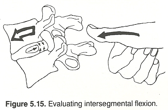
Discussion Several integrative approaches emerged, from Rome’s 296 synonyms to Bergman’s Pain/Tenderness, Asymmetry/Alignment, Range of Motion Abnormality, Tissue Tone, Texture, Temperature Abnormality, and Special Tests (PARTS) analysis adopted by the profession in the United States. Other noteworthy contributions included Ruch’s Atlas of Common Subluxations, Epstein’s introduction of network spinal analysis, and Kent’s review of CVS models. Boone’s introduction of the Journal of Vertebral Subluxation Research was accompanied by his 3-part model with Dobson. These years also included the paradigm statement of the Association of Chiropractic Colleges, which was adopted by the American Chiropractic Association, International Chiropractors Association, and World Federation of Chiropractic. Two other papers included Nelson’s critique of the CVS paradigm and Keating’s 1996 “Hunt for the Subluxation.”
There are more articles like this @ our: