Support Chiropractic Research!

Radiology

MRI Findings of Disc Degeneration are More Prevalent in Adults with Low Back Pain than in Asymptomatic Controls: A Systematic Review and Meta-Analysis

By |June 26, 2024|Low Back Pain, Radiology|

MRI Findings of Disc Degeneration are More Prevalent in Adults with Low Back Pain than in Asymptomatic Controls: A Systematic Review and Meta-Analysis

The Chiro.Org Blog


SOURCE:   AJNR Am J Neuroradiol 2015 (Dec)

  OPEN ACCESS   


W. Brinjikji • F.E. Diehn • Jarvik
C.M. Carr • Murad • P.H. Luetmera

Department of Neurological Surgery and Health Services,
Comparative Effectiveness Cost and Outcomes Research Center (J.G.J.)
Department of Radiology (J.G.J.),
University of Washington,
Seattle, Washington.


Background and purpose:   Imaging features of spine degeneration are common in symptomatic and asymptomatic individuals. We compared the prevalence of MR imaging features of lumbar spine degeneration in adults 50 years of age and younger with and without self-reported low back pain.

Materials and methods:   We performed a meta-analysis of studies reporting the prevalence of degenerative lumbar spine MR imaging findings in asymptomatic and symptomatic adults 50 years of age or younger. Symptomatic individuals had axial low back pain with or without radicular symptoms. Two reviewers evaluated each article for the following outcomes: disc bulge, disc degeneration, disc extrusion, disc protrusion, annular fissures, Modic 1 changes, any Modic changes, central canal stenosis, spondylolisthesis, and spondylolysis. The meta-analysis was performed by using a random-effects model.

There is more like this @ our

RADIOLOGY Section and the

LOW BACK PAIN Section

(more…)

Examining Clinical Opinion and Experience Regarding Utilization of Plain Radiography of the Spine: Evidence from Surveying the Chiropractic Profession

By |March 29, 2023|Chiropractic Care, Radiology|

Examining Clinical Opinion and Experience Regarding Utilization of Plain Radiography of the Spine: Evidence from Surveying the Chiropractic Profession

The Chiro.Org Blog


SOURCE:   J Clinical Medicine 2023 (Mar 10); 12 (6): 2169

  OPEN ACCESS   

Philip A Arnone, Steven J Kraus, Derek Farmen, Douglas F Lightstone, Jason Jaeger, Christine Theodossis

Community Based Internship Program, Associate Faculty,
Southern California University of Health Sciences,
Whittier, CA 90604, USA.


FROM:   Umeå University, Sweden 2008


Plain Radiography of the spine (PROTS) is utilized in many forms of healthcare including the chiropractic profession; however, the literature reflects conflicting opinions regarding utilization and value. Despite being an essential part of Evidence-Based Practice (EBP), few studies assess Doctors of Chiropractic (DCs) clinical opinions and experience regarding the utilization of (PROTS) in practice. In this study, DCs were surveyed regarding utilization of PROTS in practice. The survey was administered to an estimated 50,000 licensed DCs by email. A total of 4,301 surveys were completed, of which 3,641 were United States (US) DCs. The Clinician Opinion and Experience on Chiropractic Radiography (COECR) scale was designed to analyze survey responses. This valid and reliable scale demonstrated good internal consistency using confirmatory factor analysis and the Rasch model.

There are more articles like this @ our:

RADIOLOGY Section

(more…)

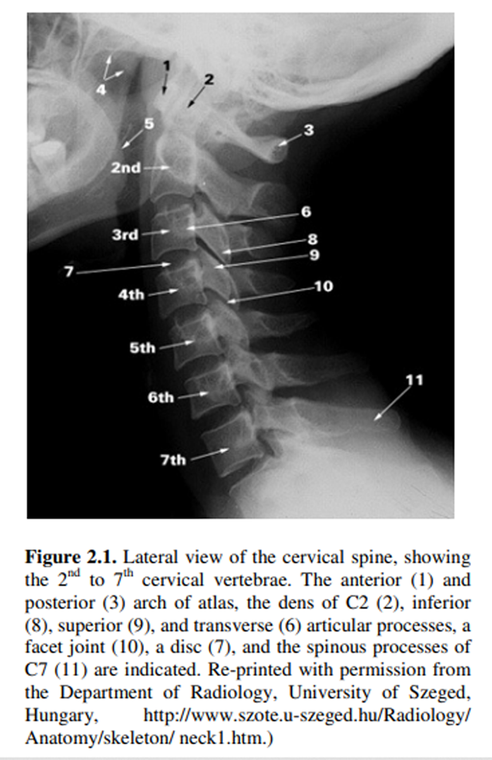
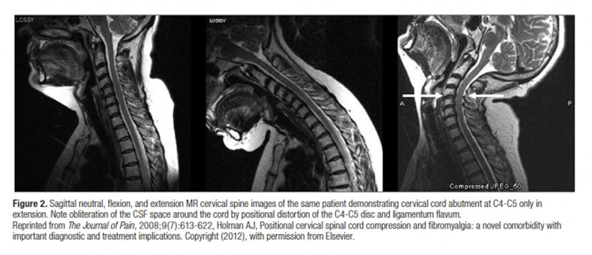
Prevalence of MRI Findings in the Cervical Spine

By |March 10, 2019|Degenerative Joint Disease, Radiology|

Prevalence of MRI Findings in the Cervical Spine in Patients with Persistent Neck Pain Based on Quantification of Narrative MRI Reports

The Chiro.Org Blog


SOURCE:   Chiropractic & Manual Therapies 2019 (Mar 6); 27: 13

Rikke Krüger Jensen, Tue Secher Jensen, Søren Grøn, Erik Frafjord, Uffe Bundgaard, Anders Lynge Damsgaard, Jeppe Mølgaard Mathiasen and Per Kjaer

Nordic Institute of Chiropractic and Clinical Biomechanics,
Odense, Denmark


BACKGROUND   Previous studies of patients with neck pain have reported a high variability in prevalence of MRI findings of disc degeneration, disc herniation etc. This is most likely due to small and heterogenous study populations. Reasons for only including small study samples could be the high cost and time-consuming procedures of having radiologists coding the MRIs. Other methods for extracting reliable imaging data should therefore be explored.

The objectives of this study were


1)   to examine inter-rater reliability among a group of chiropractic master students in extracting information
about cervical MRI-findings from radiologists´ narrative reports, and

2)   to describe the prevalence of MRI findings in the cervical spine among different age groups in patients above
age 18 with neck pain.

METHOD   Adult patients with neck pain (with or without arm pain) seen in a public hospital department between 2011 and 2014 who had an MRI of the cervical spine were identified in the patient registry ‘SpineData’. MRI-findings were extracted and quantified from radiologists’ narrative reports by second-year chiropractic master students based on a set of coding rules for the process.

The inter-rater reliability was quantified with Kappa statistics and the prevalence of the MRI findings were calculated.

RESULTS   In total, narrative MRI reports from 611 patients were included. The patients had a mean age of 52 years (SD 13; range 19–87) and 63% were women. The inter-observer agreement in coding MRI findings ranged from substantial (κ = 0.78, CI: 0.33–1.00) to almost perfect (κ = 0.98, CI: 0.95–1.00).

The most prevalent MRI findings were foraminal stenosis (77%), uncovertebral arthrosis (74%) and disc degeneration (67%) while the least prevalent findings were nerve root compromise (2%) and Modic changes type 2 (6%). Modic type 1 was mentioned in 25% of the radiologists’ reports. The prevalence of all findings increased with age, except disc herniation which was most prevalent for patients in their forties.

There are more articles like this @ our:

RADIOLOGY Section

(more…)

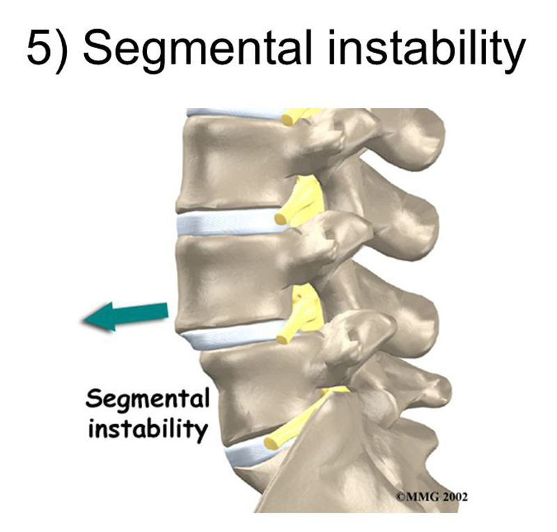
Kinematic Analysis of Dynamic Lumbar Motion in Patients

By |January 23, 2019|Radiology|

Kinematic Analysis of Dynamic Lumbar Motion in Patients with Lumbar Segmental Instability Using Digital Videofluoroscopy

The Chiro.Org Blog


SOURCE:   Eur Spine J. 2009 (Nov); 18 (11): 1677–1685

Amir Ahmadi, Nader Maroufi, Hamid Behtash, Hajar Zekavat, and Mohamad Parnianpour

Faculty of Rehabilitation,
Iran University of Medical Sciences,
P.O. Box 15875-4391,
Tehran, Iran.


The study design is a prospective, case-control. The aim of this study was to develop a reliable measurement technique for the assessment of lumbar spine kinematics using digital video fluoroscopy in a group of patients with low back pain (LBP) and a control group. Lumbar segmental instability (LSI) is one subgroup of nonspecific LBP the diagnosis of which has not been clarified. The diagnosis of LSI has traditionally relied on the use of lateral functional (flexion-extension) radiographs but use of this method has proven unsatisfactory. Fifteen patients with chronic low back pain suspected to have LSI and 15 matched healthy subjects were recruited. Pulsed digital videofluoroscopy was used to investigate kinematics of lumbar motion segments during flexion and extension movements in vivo. Intersegmental linear translation and angular displacement, and pathway of instantaneous center of rotation (PICR) were calculated for each lumbar motion segment. Movement pattern of lumbar spine between two groups and during the full sagittal plane range of motion were analyzed using ANOVA with repeated measures design.

Intersegmental linear translation was significantly higher in patients during both flexion and extension movements at L5-S1 segment (p < 0.05). Arc length of PICR was significantly higher in patients for L1-L2 and L5-S1 motion segments during extension movement (p < 0.05). This study determined some kinematic differences between two groups during the full range of lumbar spine. Devices, such as digital videofluoroscopy can assist in identifying better criteria for diagnosis of LSI in otherwise nonspecific low back pain patients in hope of providing more specific treatment.

There are more articles like this @ our:

RADIOLOGY Section

(more…)

Injuries in the Pediatric Patient

By |December 25, 2015|Pediatrics, Radiology|

Injuries in the Pediatric Patient: Review of Key Acquired and Developmental Issues

The Chiro.Org Blog


J Clin Chiro Peds 2009 (Dec); 10 (2): 665—670 ~ FULL TEXT


Beverly L. Harger, D.C., D.A.C.B.R., and Kim Mullen, D.C.

Department of Radiology,
Western States Chiropractic College,
Portland, Oregon


Key words:   pediatric trauma, growth plate fractures, slipped capital femoral epiphysis, incomplete fractures, torus fracture, spondylolysis, pars interarcularis defects, single photon emission computed tomography, Scheuermann’s disease, ring apophyseal fracture.


Introduction

A plethora of conditions specifically target children and adolescents which are not prevalent in the adult population. Understanding the age-related differences in this population can help clinicians improve diagnosis and therefore management of these conditions. Though it is beyond the scope of this paper to extensively address diseases targeting the pediatric population, common key injuries will be discussed with emphasis on the role imaging plays in establishing accurate diagnosis.

There are more articles like this @ our:

Chiropractic Pediatrics Page and the:

Chiropractic Radiology Page

(more…)

Radiology Undergraduate and Resident Curricula

By |December 12, 2015|Radiology|

Radiology Undergraduate and Resident Curricula: A Narrative Review of the Literature

The Chiro.Org Blog


SOURCE:   J Chiropractic Humanities 2015 (Dec); (22) 1: 1–8 ~ FULL TEXT


Kathleen L. Linaker, DC, DACBR, PhD

Dean, Center for Life and Health Sciences,
Mohawk Valley Community College,
Utica, NY


Objective   The purpose of this study was to examine the literature regarding radiology curricula for both undergraduates and residents.

Methods   A review of the literature was performed using relevant key words. Articles were retrieved through December 2012 using PubMed, ScienceDirect, ERIC, Proquest, and ICL databases along with a manual review of references.

Results   Of the 4716 unique abstracts reviewed by the author, 142 were found to be relevant to the purpose of this study. Undergraduate radiology education, radiology curriculum, and radiology pedagogy vary widely between disciplines and between colleges within disciplines. Formal radiology education is not taught at all medical programs and little radiology training is incorporated into non-radiology residencies. This results in some medical graduates not being taught how to interpret basic radiology images and not learning contraindications and indications for ordering diagnostic imaging tests. There are no definitive studies examining how to incorporate radiology into the curriculum, how to teach radiology to either undergraduates or residents, or how to assess this clinical competency.

There are more articles like this @ our:

Chiropractic Radiology Section

(more…)