
Chiropractic Technique Review: Pierce Results System
SOURCE: Chiro.Org’s Technique Page
The Chiropractic Technique, known as the Pierce Results System, (formerly known as Pierce/Stillwagon, or PST) was developed by Vernon Pierce, D.C., Sr. It is a biomechanical analysis of spine kinematics (or spinal motion), utilizing “stress views” of the spine (flexion, extension, rotation, and/or lateral bending views where required) or videofluoroscopy (VF, or “moving x-ray” studies) to determine the loss of spinal function, which is at the core of the “vertebral subluxation complex”.
Structure and Function
Our spine is a “structural” unit. There are 4 curves to the spine. Loss of structural integrity and/or normal function of the spine is the basis for the evolution of the vertebral subluxation. Abnormal stresses occur in the facets, discs and supporting tissues when normal motion of the spine is impaired. The Pierce System analysis is aimed at locating the specific segments which are subluxated, as well as providing the means to “free” those segments.
When normal function returns, the neurologic and other components of the subluxation complex resolve by the normal healing power referred to historically as “innate intelligence” or “vis medicatrix naturae” (the healing power of Nature).
| Neutral Lateral Film Analysis: |
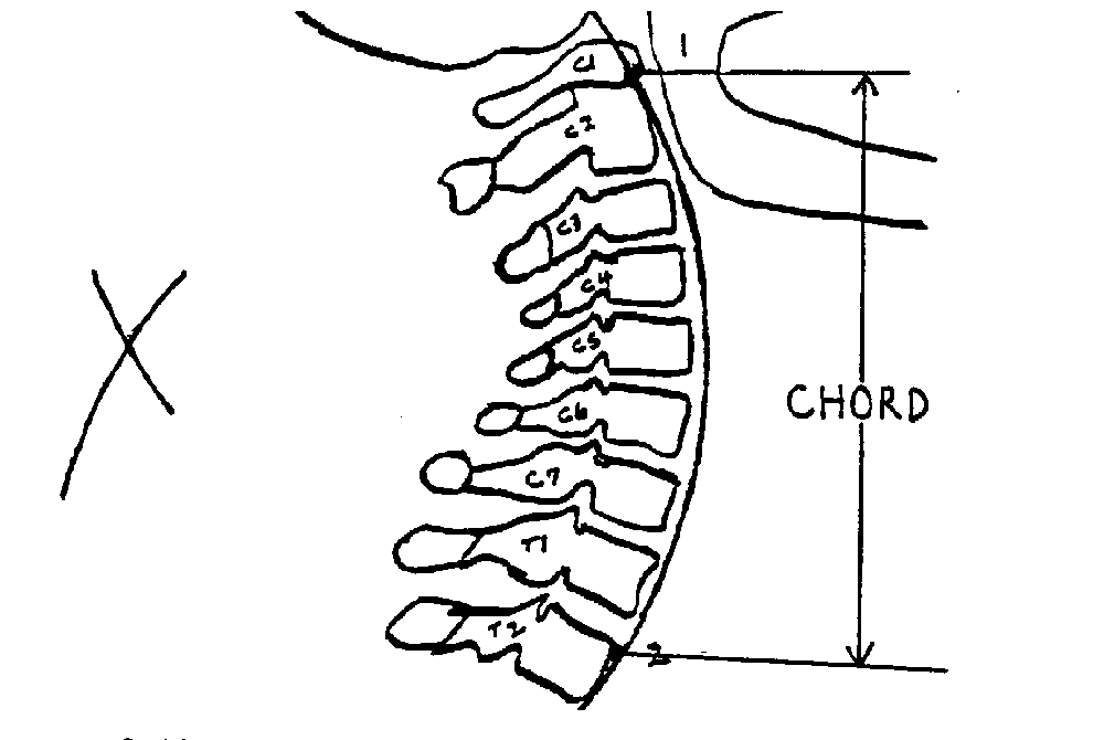 |
The picture on the left is an example of perfect cervical lordosis. All segments should be on Georges’s line (one curved line). There should be an even spacing between each spinous process. Positioning of the head and spine should also be assessed for anterior head placement (also known as Forward Head Posture). The normal cervical lordosis (which extends from C1 to T2) should have a 17-24 cm. radius, based on the patient’s height. This is easily measured with the AcuArc ruler. The posterior arch of Atlas should be centered in the space between occiput and the C2 spinous process. If C1’s posterior arch “crowds” occiput, it is labelled as an “inferior” Atlas. If it crowds C2, it is labelled “superior”. The normal Atlas Plane line would be 18-24 degrees superior to the bottom of the film. A line under the bottom of the C2 body (Whitehorn’s line) should be parallel with the floor.
| Flexion Film Analysis: |
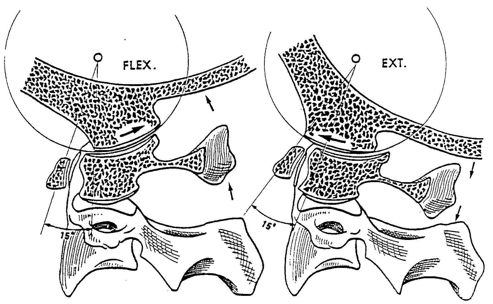 |
NOTE: When pre-positioning the patient for this view, it’s KEY to have them first lower their chin before they flex their spine, otherwise you may not observe motion at occiput. The picture on the left demonstrates “nutation” at occiput. Occiput reproduced with permission from Physiology of the Joints, Volume III |
 |
The picture on the left is an example of perfect cervical flexion. When the spine flexes, it should fully reverse the cervical curve. Three primary motions should occur in flexion:
(1) The zygapophyses (facets) should slide upwards and forwards. Because of this motion,
(2) the IVF’s should open (more) fully. And lastly,
(3) the spinous processes should “fan out”, or separate.
Occiput should flex forwards, and the posterior arch of C1 should approximate the back of occiput.
In flexion, George’s (posterior body) line should be one curved line, and all segments should be on that line. If you require more than one line to connect all the segments, the subluxated segment will reside in the portion of the spine which is straightened. In flexion lock, typically the segment just below the intersection of these 2 lines is the subluxated segment. The most obvious indicator would be that the segment fails to flex and thus, increase the size of it’s IVF.
| Extension Film Analysis: |
You can learn much more about the Pierce analysis at our:
I’ve been doing motion studies on cervical spines and it is amazing to see how many people will have 3 to 4 levels that will not flex or extend.
Dr. Adam
Certainly, in flexion, if C5 fails to flex, most often, all the segments below C5 won’t move much either.
That’s where Dr. Pierces flexion rule comes in handy.
When it takes 2 lines to connect the vertebra, it’s usually the vertebra JUST BELOW the intersection of these lines that is the problem issue.
If you adjust THAT segment, and then visualized them in flexion again (and I am not recommending that…I am reporting what was seen on hundreds of pre- and post- adjustment VF studies in the 90s) you would find that ALL the segments below that kink were moving again.
I guess that’s a subtle way of saying you don’t have to mess with all of them, just the ONE that was causing the loss of motion. This is the classic case of “less is more”.
Do you ave a source so I can get my hands on the rules?
Thanks!
Hi David
The Pierce Page [http://www.chiro.org/ChiroZine/pierce.shtml] covers most of the didactic information. There are also Pierce Results classes being offered near Life College (Marietta) and occasionally around the country. Check with their website (as listed on my Pierce Page)