Motion Palpation of the Cervical Spine
We would all like to thank Dr. Richard C. Schafer, DC, PhD, FICC for his lifetime commitment to the profession. In the future we will continue to add materials from RC’s copyrighted books for your use.
This is Chapter 3 from RC’s best-selling book:
“Motion Palpation”
These materials are provided as a service to our profession. There is no charge for individuals to copy and file these materials. However, they cannot be sold or used in any group or commercial venture without written permission from ACAPress.
Chapter 3: The Cervical Spine
This chapter describes the basic biomechanical, diagnostic, and therapeutic considerations related to motion palpation and the cervical spine. Emphasis will be on relating the general concepts previously explained about the chiropractic fixation-subluxation complex to specific entities that can be revealed by motion palpation and frequently corrected by dynamic chiropractic. Some aids to differential diagnosis are also included.
APPLIED ANATOMY CONSIDERATIONS
There are seven sites of possible “articular” fixation in the cervical spine. They are at the bilateral apophyseal joints, the bilateral covertebral joints, the superior and inferior intervertebral disc (IVD) interfaces, and the odontal-atlantal articulation (Table 3.1).
Table 3.1. The 27 Sites of Possible Spinopelvic Articular Fixation
| In the cervical spine (7 possible sites of fixation) | |
| Bilateral apophyseal joints | |
| Bilateral covertebral joints | |
| Superior and inferior IVD interfaces | |
| Odontal-atlantal articulation | |
| In the thoracic spine (8 possible sites of fixation) | |
| Bilateral apophyseal joints | |
| Superior and inferior IVD interfaces | |
| Bilateral costovertebral joints | |
| Bilateral costotransverse joints | |
| In the lumbar spine (4 possible sites of fixation) | |
| Bilateral apophyseal joints | |
| Superior and inferior IVD interfaces | |
| In the pelvis (8 possible sites of fixation) | |
| Bilateral superior sacroiliac joints | |
| Bilateral inferior sacroiliac joints | |
| Sacrococcygeal joint | |
| Pubic joint | |
| Bilateral acetabulofemoral joints | |
The Apophyseal Joints of the Spine
Throughout the spine, paired diarthrodial articular processes (zygapophyses) project from the vertebral arches. The superior processes (prezygapophyses) of the inferior vertebra contain articulating facets that face somewhat posteriorly. They mate with the inferior processes (postzygapophyses) of the vertebra above that face somewhat anteriorly. Each articular facet is covered by a layer of hyaline cartilage that faces the synovial joint. The angulation of vertebral facets normally varies with the level of the spine and can be altered by wear and pathology.
In visualizing the motion of any joint, it is helpful to keep in mind that the hyaline-coated articulating surface is not the shape of the often flat bony surface exhibited on an x-ray film. Most apophyseal joints of the spine have a convex-concave shape.
Fisk states that the posterior joints of the spine are more prone to osteoarthritic changes than any other joint in the body: “Evidence of disc degeneration precedes this arthritis in the lumbar spine, but there is no such relationship in the cervical spine.” However, most authorities agree with Grieve that the presence of arthrotic changes in the facet planes does not, of itself, necessarily have any effect on ranges of movement, neither does the presence of osteophytosis.
Regional Structural Characteristics
| Review the complete Chapter (including sketches and Tables) at the ACAPress website |


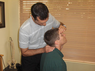
I palpate the neck sitting or prone, or supine. How come some people you dont even have to motion their neck and it feels like their C1 or C2 is coming out of their neck? Thanks for the great information, and when I was in Chiro school I enjoyed gonstead because all the motion palpation and study of the biomechanics we got.
Marco, D.C.