Support Chiropractic Research!

Radiology

Radiologic Manifestations of Spinal Subluxations

By |March 30, 2012|Chiropractic Care, Radiology, Subluxation|

Radiologic Manifestations of Spinal Subluxations

The Chiro.Org Blog


We would all like to thank Dr. Richard C. Schafer, DC, PhD, FICC for his lifetime commitment to the profession. In the future we will continue to add materials from RC’s copyrighted books for your use.

This is Chapter 6 from RC’s best-selling book:
“Basic Chiropractic Procedural Manual”

These materials are provided as a service to our profession. There is no charge for individuals to copy and file these materials. However, they cannot be sold or used in any group or commercial venture without written permission from ACAPress.


Chapter 6: Radiologic Manifestations of Spinal Subluxations

This chapter describes the radiologic signs that may be expected when spinal subluxations are demonstrable by radiography. Through the years, there have been several concepts within the chiropractic profession about what actually constitutes a subluxation. Each has had its rationale (anatomical, neurologic, or kinematic), and each has had certain validity contributing to our understanding of this complex phenomenon.

You may review the full Chapter 6 @:

Radiologic Manifestations of Spinal Subluxations


Kinetic Intersegmental Subluxations


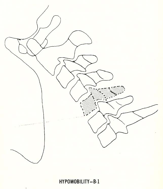
Segmental hypomobility, also called a “fixation subluxation” by many clinicians, may affect one or several motor units.

It is characterized by reduced motion of the “Spinal Motion Unit” (Please refer to Spinal Anatomy 101), which has been forced to the extreme of a range of motion (eg, flexion, extension, etc). See Figure 6.14. Stress views or videofluoroscopy are necessary to depict this and other kinetic subluxations radiographically, but motion palpation and some orthopedic tests may reveal their presence clinically.

Editor’s Note:   In the following picture, the inferior facet of C5 fails to slide forwards and upwards upon the superior facet of C6. Because of that, the IVF cannot open more fully, and the spinous process of C5 fails to move away from the C6 spinous. All together, these are the classic signs of HYPO-mobility.

(more…)

MRI Update

By |July 9, 2011|Radiology|

MRI Update

The Chiro.Org Blog


SOURCE:  ACA News

By Caitlin Lukacs


Technological progress is not reserved for cell phones and iPads. Advanced imaging modalities, such as magnetic resonance imaging (MRI), have benefited from recent advances as well. In fact, MRI has become the gold standard of advanced imaging for the spine and extremity joints. “It’s hard to do anything in medicine without imaging,” says Norman Kettner, DC, DACBR, professor of clinical science and chairman of the Department of Radiology at Logan College of Chiropractic. “In fact, in a survey of the top 25 internists in the United States, MRI and CT scans were rated No. 1 as the most important development in medical science in the previous century.

What Is MRI?

Gary Longmuir, DC, DACBR, clinical radiologist and president of the ACA Council on Diagnostic Imaging, explains that MRIs create images of the internal structures of the body based on the energy released from hydrogen protons. Because the body is largely made up of water and each water molecule contains two hydrogen protons, when a person enters the magnetic field created by the MRI scanner, the magnetic charges of the protons change and align with the direction of the scanner’s field. When the field is then turned off, the protons return to their original energy state, releasing the energy difference as photons that are detected by the scanner as a “signal” similar to a radio wave. The protons of distinct tissues return to their original energy states at different rates, and this difference is detected by the MRI scanner.

DCs and MRI (more…)

The Pediatric Elbow: A Review of Fractures

By |June 18, 2011|Pediatrics, Radiology|

The Pediatric Elbow: A Review of Fractures

The Chiro.Org Blog


SOURCE:   Dynamic Chiropractic

By Deborah Pate, DC, DACBR


The elbow fracture is one of the most common fractures in children. Assessing the elbow for fracture can be difficult because of the changing anatomy of the growing skeleton and the subtlety of some of these fractures.

It’s important to be aware of the radiographic signs of fracture in the elbow, along with knowing the appearance and fusion of the ossification centers in the pediatric patient, to avoid confusing an ossification center with a fracture fragment. Of course, alignment and radiographic positioning are also extremely important in making a diagnostic assessment.


Diagram of a distended joint capsule
with the fat pads displaced.

When reviewing an X-ray study for trauma, it’s best to have a methodical way of viewing the study. Perform (at the very least) two views of the elbow at 90° to each other, AP and lateral. Positioning is very important, particularly in the growing skeleton. Alignment of the joint cannot be assessed unless the positioning is accurate.

The two most common errors in positioning are: 1) elbow is lower than the shoulder, which projects the capitellum onto the ulna; and 2) elbow is higher than the wrist, which will make the capitellum and the head of the radius appear anterior, and the epicondyle appear posterior, making it difficult to assess the alignment. In a true lateral view, the elbow and the shoulder should be in the same plane and the wrist should be higher than the elbow to compensate for the normal valgus position of the elbow. The thumb should be up to keep the radius from rotating. (more…)