

Support Chiropractic Research!

Chiropractic Care

Do Participants With Low Back Pain Who Respond to Spinal Manipulative Therapy Differ Biomechanically?

By |September 1, 2015|Chiropractic Care, Low Back Pain, Spinal Manipulation|

Do Participants With Low Back Pain Who Respond to Spinal Manipulative Therapy Differ Biomechanically From Nonresponders, Untreated Controls or Asymptomatic Controls?

The Chiro.Org Blog


SOURCE:   Spine 2015 (Sep 1);   40 (17):   1329–1337 ~ FULL TEXT


Wong, Arnold Y. L. PT, MPhil, PhD; Parent, Eric C. PT, PhD;
Dhillon, Sukhvinder S. MB, ChB, CCST; Prasad, Narasimha PhD;
Kawchuk, Gregory N. DC, PhD

Department of Rehabilitation Sciences,
The Hong Kong Polytechnic University,
Kowloon, Hong Kong

Department of Physical Therapy,
University of Alberta,
Alberta, Canada


FROM:  University of Alberta ~ 8-31-2015

Researchers at the University of Alberta have found that spinal manipulation—applying force to move joints to treat pain, a technique most often used by chiropractors and physical therapists — does indeed have immediate benefits for some patients with low-back pain but does not work for others with low-back pain. And though on the surface this latest conflict might appear to muddy the waters further, the results point to the complexity of low-back pain and the need to treat patients differently, says lead author Greg Kawchuk.“This study shows that, just like some people respond differently to a specific medication, there are different groups of people who respond differently to spinal manipulation.”

In a non-randomized control study, individuals with low-back pain received spinal manipulation during two treatment sessions that spanned a week. Participants reported their pain levels and disability levels after spinal manipulation, and researchers used ultrasound, MRI and other diagnostics to measure changes in each participant’s back, including muscle activity, properties within the intervertebral discs, and spinal stiffness.

A control group of participants with low-back pain underwent similar clinical examinations but did not receive spinal manipulation. A third group — those who did not have low-back pain symptoms — were also evaluated.

The people who responded to spinal manipulation reported less pain right away and showed improvement in back muscle thickness, disc diffusion and spinal stiffness. Those changes were great enough to exceed or equal the measures in the control groups and stayed that way for the week of treatment, the research team found.
A patient receives spinal manipulation treatment.

Kawchuk, who practised as a chiropractor before going on to obtain his PhD in biomechanics and bioengineering, said the results do not advocate one way or another for spinal manipulation but help explain why there has been so much conflicting data about its merits.

“Clearly there are some people with a specific type of back pain who are responding to this treatment and there are some people with another type of back pain who do not. But if you don’t know that and you mix those two groups together, you get an artificial average that doesn’t mean anything,” Kawchuk explained.

The research team is still fine-tuning how to distinguish who is a responder or non-responder before spinal manipulation is given; however, this study shows it can be used to identify an effective treatment course.

“Spinal manipulation acts so rapidly in responders that it could be used as a screening tool to help get the right treatment to the right patient at the right time.”

The study did not investigate the long-term effects of spinal manipulation, but this is next on the list for the researchers.


STUDY DESIGN:   Nonrandomized controlled study.

OBJECTIVE:   To determine whether patients with low back pain (LBP) who respond to spinal manipulative therapy (SMT) differ biomechanically from nonresponders, untreated controls or asymptomatic controls.

SUMMARY OF BACKGROUND DATA:   Some but not all patients with LBP report improvement in function after SMT. When compared with nonresponders, studies suggest that SMT responders demonstrate significant changes in spinal stiffness, muscle contraction, and disc diffusion. Unfortunately, the significance of these observations remains uncertain given methodological differences between studies including a lack of controls.

There are more articles like this @ our:
Low Back Pain and Chiropractic Page

(more…)

Craniocervical Chiropractic Procedures – A Précis of Upper Cervical Chiropractic

By |August 9, 2015|Chiropractic Care, Upper Cervical Adjusting|

Craniocervical Chiropractic Procedures – A Précis of Upper Cervical Chiropractic

The Chiro.Org Blog


SOURCE:   J Can Chiropr Assoc 2015 (Jun);   59 (2):   173–192 ~ FULL TEXT


H. Charles Woodfield, III, BPhm, DC, Craig York, DC.
Roderic P. Rochester, DC, Scott Bales, DC,
Mychal Beebe, DC, Bryan Salminen, DC,
Jeffrey N. Scholten, DC .

Director of Research –
Upper Cervical Research Foundation,
Minneapolis, MN


Presented here is a narrative review of upper cervical procedures intended to facilitate understanding and to increase knowledge of upper cervical chiropractic care. Safety, efficacy, common misconceptions, and research are discussed, allowing practitioners, chiropractic students, and the general public to make informed decisions regarding utilization and referrals for this distinctive type of chiropractic care. Upper cervical techniques share the same theoretical paradigm in that the primary subluxation exists in the upper cervical spine. These procedures use similar assessments to determine if spinal intervention is necessary and successful once delivered. The major difference involves their use of either an articular or orthogonal radiograph analysis model when determining the presence of a misalignment. Adverse events following an upper cervical adjustment consist of mild symptomatic reactions of short-duration (< 24-hours). Due to a lack of quality and indexed references, information contained herein is limited by the significance of literature cited, which included non-indexed and/or non-peer reviewed sources.

KEYWORDS:   adverse events; atlas; cervical; chiropractic; chiropractic adjustment; craniocervical


From the Full-Text Article:

Introduction:

The indexed literature reports the existence of many upper cervical (UC) procedures. [1, 2] Presented here is a brief narrative review or narrative description of upper cervical techniques (UCT) with the intention of increasing knowledge and understanding regarding their effectiveness and utilization. Procedural similarities and differences between UCT are examined. As chiropractic goes the way of other healing professions through stratification into specialties, this characterization of upper cervical procedures can create appreciation and clarity both inside and outside the profession.

There are more articles like this @ our:

Chiropractic Technique Page

(more…)

What Effect Does Chiropractic Treatment Have on Gastrointestinal (GI) Disorders

By |August 6, 2015|Chiropractic Care, Gastrointestinal Disorders, Visceral Disease|

What Effect Does Chiropractic Treatment Have on Gastrointestinal (GI) Disorders: A Narrative Review of the Literature

The Chiro.Org Blog


SOURCE:   J Can Chiropr Assoc 2015 (Jun);   59 (2):   122–133 ~ FULL TEXT


Katherine Angus, BSc(Kin), DC, Sepideh Asgharifar, BSc(Hons), DC,
Brian Gleberzon, DC, MHSc

Chiropractic and Health Associates Clinic,
1 St. Clair Ave West, Suite 404,
Toronto, Ontario.


The purpose of this study was to provide a narrative review of the literature of studies describing the management of disorders of the gastro-intestinal (GI) tract using ‘chiropractic therapy’ broadly defined here as spinal manipulation therapy, mobilizations, soft tissue therapy, modalities and stretches. Search limiters include access to full text studies published between 1980 and November 2012 in peer-reviewed journals, English language only involving human subjects. Twenty-one articles were found that met our inclusion criteria. Retrievable articles varied from case reports to clinical trials to review articles of management options. The majority of articles chronicling patient experiences under chiropractic care reported they demonstrated mild to moderate improvements in presenting symptoms. No adverse side effects were reported. This suggests chiropractic care can be considered as an adjunctive therapy for patients with various GI conditions providing there are no co-morbidities.

KEYWORDS:  chiropractic; gastro-intestinal; manipulation; therapy


From the FULL TEXT Article

Introduction

The purpose of this paper was to conduct a narrative review of the literature that investigated the effectiveness of chiropractic treatment for gastrointestinal (GI) disorders. A previous review by Gleberzon et al [1] reviewed the literature from 2007 to 2011 that investigated the use of one type of chiropractic treatment (spinal manipulative therapy) for pediatric health conditions one of which was colic, often considered a type of GI condition effecting infants [Authors’ note: there is considerable debate whether or not ‘infantile colic’ is a GI condition, or simply ‘baby back pain’ and there is debate if the method used to monitor its existence (crying time) is a subjective or objective outcome measure. [1] That debate notwithstanding, for the purposes of this report, we have included colic as a GI condition].

There are more articles like this @ our:

Conditions That Respond Well to Chiropractic Page

(more…)

Back Pain in Adolescents With Idiopathic Scoliosis

By |June 30, 2015|Chiropractic Care, Scoliosis|

Back Pain in Adolescents With Idiopathic Scoliosis: Epidemiological Study for 43,630 Pupils in Niigata City, Japan

The Chiro.Org Blog


SOURCE:   Eur Spine J. 2011 (Feb);   20 (2):   274–279 ~ FULL TEXT


Tsuyoshi Sato, Toru Hirano, Takui Ito, Osamu Morita, Ren Kikuchi,
Naoto Endo, and Naohito Tanabe

Department of Orthopedic Surgery,
Niigata Prefectural Shibata Hospital,
Shibata, Japan.
tsuyoshis1@mac.com


There have been a few studies regarding detail of back pain in adolescents with idiopathic scoliosis (IS) as prevalence, location, and severity. The condition of back pain in adolescents with IS was clarified based on a cross-sectional study using a questionnaire survey, targeting a total of 43,630 pupils, including all elementary school pupils from the fourth to sixth grade (21,893 pupils) and all junior high pupils from the first to third year (21,737 pupils) in Niigata City (population of 785,067), Japan.

32,134 pupils were determined to have valid responses (valid response rate: 73.7%). In Niigata City, pupils from the fourth grade of elementary school to the third year of junior high school are screened for scoliosis every year. This screening system involves a three-step survey, and the third step of the survey is an imaging and medical examination at the Niigata University Hospital.

In this study, the pupils who answered in the questionnaire that they had been advised to visit Niigata University Hospital after the school screening were defined as Scoliosis group (51 pupils; 0.159%) and the others were defined as No scoliosis group (32,083 pupils). The point and lifetime prevalence of back pain, the duration, the recurrence, the severity and the location of back pain were compared between these groups.

The severity of back pain was divided into three levels (level 1 no limitation in any activity; level 2 necessary to refrain from participating in sports and physical activities, and level 3 necessary to be absent from school). The point prevalence was 11.4% in No scoliosis group, and 27.5% in Scoliosis group. The lifetime prevalence was 32.9% in No scoliosis group, and 58.8% in Scoliosis group. According to the gender- and school-grade-adjusted odds ratios (OR), Scoliosis group showed a more than twofold elevated odds of back pain compared to No scoliosis group irrespective of the point or lifetime prevalence of back pain (OR, 2.29; P = 0.009 and OR, 2.10; P = 0.012, respectively).

Scoliosis group experienced significantly more severe pain, and of a significantly longer duration with more frequent recurrences in comparison to No scoliosis group. Scoliosis group showed significantly more back pain in the upper and middle right back in comparison to No scoliosis group. These findings suggest that there is a relationship between pain around the right scapula in Scoliosis group and the right rib hump that is common in IS.


From the Full-Text Article:

Introduction:

Most patients with adolescent idiopathic scoliosis (AIS) visit the hospital when a trunk deformity, such as rib or lumbar hump and waist asymmetry, is pointed out either after the school screening or by family members, and it is rare for these patients to visit the hospital due to back pain. However, some adolescent patients with idiopathic scoliosis (IS) do complaint of back pain in outpatient clinics. Previously, it had been accepted that special attention should be paid to patients with scoliosis who experienced back pain, because it was thought that might be additional pathologies such as an occult syrinx, spinal cord tumors, or neuromuscular disorders [4, 6, 20].

There are more articles like this @ our:

Scoliosis and Chiropractic Page

(more…)

DCs Treating the Multiple Sclerosis Patient

By |May 29, 2015|Chiropractic Care, Multiple Sclerosis|

DCs Treating the Multiple Sclerosis Patient

The Chiro.Org Blog


SOURCE:   ACA News ~ May 2015 ~ FULL TEXT


By Lori A. Burkhart


Multiple Sclerosis (MS) is the most common disabling neurological disease of young adults, according to the National Institutes of Health (NIH), most often appearing when people are between 20 and 40 years old. However, it can also affect children and adults over 40. The U.S. National Library of Medicine defines MS as an autoimmune disease that affects the central nervous system (brain and spinal cord). The myelin sheath, a protective membrane that wraps around the axon of a nerve cell, is destroyed in a patient with MS; this is caused by inflammation. That damage causes nerve signals to slow down or stop. MS affects women more than men.

Since doctors of chiropractic are recognized as primary contact neuromusculoskeletal specialists, most will have patients with undiagnosed MS come into their practices. The DC will diagnose the patient, treat certain symptoms and make the appropriate referrals.


Diagnosis

Diagnosis of MS is complicated in that it can be severe or mild and can go into remission. NIH points out that initial symptoms often are double or blurred vision, red-green color distortion or blindness in one eye. Most MS patients experience muscle weakness in their extremities and difficulty with coordination and balance.

According to Larry Wyatt, DC, DACBR, FICC, professor and senior faculty, division of clinical sciences at Texas Chiropractic College, MS is diagnosed in a number of ways, as its clinical course is distinctive in each patient and there are different types of MS. Some patients with obvious MS are diagnosed by clinical signs and symptoms (i.e., attacks) alone. These patients will have MS attacks that often relapse for months or even years. In other patients further testing is necessary. Magnetic resonance imaging (MRI), often with gadolinium enhancement, is the mainstay of diagnosis in most cases. “Patients with MS will very often have multiple high-signal intensity lesions in the brain and/or spinal cord on T2-weighted images,” Dr. Wyatt says. “In addition, cerebrospinal fluid analysis for immunoglobulin content can be quite helpful. There is a specific set of criteria, called the McDonald Criteria, which outline the findings necessary for the diagnosis of the different forms of MS.”

Jason West, DC, DCBCN, a fourth-generation DC who operates a clinic in Pocatello, Idaho, says the majority of the diagnosis comes from the patient history, but he points out that usually when patients with MS come in, they already are diagnosed and they are unhappy with their medical treatment options. “If they weren’t diagnosed, one of the standards is to do an MRI and look for white lesions, and there is also a spinal tap to look for antibodies,” Dr. West says. “Usually these patients have a history of peripheral neuropathy or neurological disease or processes occurring.”


Symptom Management

(more…)

Real-Time Visualization of Joint Cavitation

By |April 16, 2015|Chiropractic Care|

Real-Time Visualization of Joint Cavitation

The Chiro.Org Blog


SOURCE:   PLoS One. 2015 (Apr 15); 10 (4): e0119470


Gregory N. Kawchuk, Jerome Fryer, Jacob L. Jaremko,
Hongbo Zeng, Lindsay Rowe, Richard Thompson

Department of Physical Therapy,
Faculty of Rehabilitation Medicine,
University of Alberta,
Edmonton, Alberta, Canada


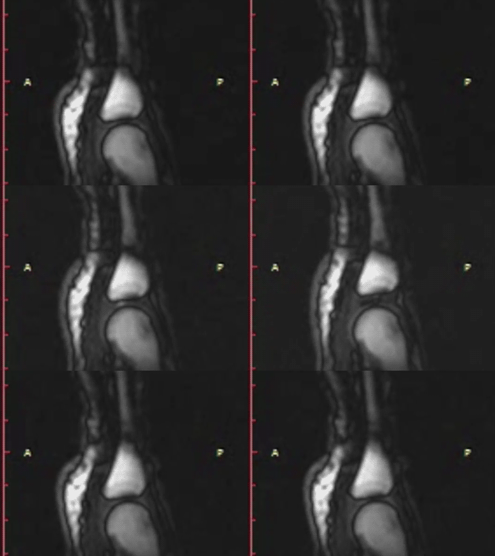
Cracking sounds emitted from human synovial joints have been attributed historically to the sudden collapse of a cavitation bubble formed as articular surfaces are separated. Unfortunately, bubble collapse as the source of joint cracking is inconsistent with many physical phenomena that define the joint cracking phenomenon. Here we present direct evidence from real-time magnetic resonance imaging that the mechanism of joint cracking is related to cavity formation rather than bubble collapse. In this study, ten metacarpophalangeal joints were studied by inserting the finger of interest into a flexible tube tightened around a length of cable used to provide long-axis traction. Before and after traction, static 3D T1-weighted magnetic resonance images were acquired. During traction, rapid cine magnetic resonance images were obtained from the joint midline at a rate of 3.2 frames per second until the cracking event occurred. As traction forces increased, real-time cine magnetic resonance imaging demonstrated rapid cavity inception at the time of joint separation and sound production after which the resulting cavity remained visible. Our results offer direct experimental evidence that joint cracking is associated with cavity inception rather than collapse of a pre-existing bubble. These observations are consistent with tribonucleation, a known process where opposing surfaces resist separation until a critical point where they then separate rapidly creating sustained gas cavities. Observed previously in vitro, this is the first in-vivo macroscopic demonstration of tribonucleation and as such, provides a new theoretical framework to investigate health outcomes associated with joint cracking.

Enjoy this live video demonstration


From the FULL TEXT Article:

Introduction

Background

Sounds emitted from human synovial joints vary in their origin. Joint sounds that occur repeatedly with ongoing joint motion arise typically when anatomic structures rub past one another. In contrast, “cracking” sounds require time to pass before they can be repeated despite ongoing joint motion. Although various hypotheses have been proposed over many decades regarding the origin of cracking sounds, none have been validated; the underlying mechanism of cracking sounds remains unknown.

History

(more…)