Support Chiropractic Research!

Diagnosis

Clinical Chiropractic: The Elbow and Forearm

By |November 14, 2011|Diagnosis, Education|

Clinical Chiropractic: The Elbow and Forearm

The Chiro.Org Blog


We would all like to thank Dr. Richard C. Schafer, DC, PhD, FICC for his lifetime commitment to the profession. In the future we will continue to add materials from RC’s copyrighted books for your use.

This is Chapter 8 from RC’s best-selling book:

“Clinical Chiropractic: Upper Body Complaints”

These materials are provided as a service to our profession. There is no charge for individuals to copy and file these materials. However, they cannot be sold or used in any group or commercial venture without written permission from ACAPress.


Chapter 8:   The Elbow and Forearm

CLINICAL BRIEFING

Functional Considerations

The arm and forearm are joined by a joint that serves as both a hinge and a pivot. The semilunar notch of the ulna is hinged with the hyperboloid trochlea of the humerus. The proximal head of the radius pivots with the spherical capitulum of the humerus and glides against both the proximal and distal ends of the ulna.

The distal end of the humerus can be viewed as two columns: a larger one medially that articulates with the semilunar notch of the ulna, and a smaller one laterally that articulates with the head of the radius. The pulley-like trochlea apparatus has:

(1) a depression at the front that lodges the coronoid process of the ulna and

(2) a depression at the rear that holds the olecranon process of the ulna when the elbow is extended.

The olecranon process restricts hyperextension of the elbow and protects the ulnohumeral articulation posteriorly. The concave head of the radius glides against the spherical capitulum of the humerus. The capitulum and trochlea are separated by a bony crest that fits into the opening between the proximal ulna and the radius and serves as a fixed rudder to guide elbow motion. The elbow flexors originate from the medial epicondyle, and the extensors originate from the lateral epicondyle. This structural arrangement should be visualized during examination to discriminate normal from abnormal articular motion.

The basic range of elbow joint motion involves elbow flexion (135°) and extension (0°), and forearm supination (90°) and pronation (90°). If a motion block is found in active motion, passive motion should be checked and the type of restriction and its degree noted.

Clinical Analysis

The elbow joint was not made to be used as an organic battering ram, but it often is: purposefully in sports; by accident in falls. For this reason, the vast majority of elbow disorders has trauma as their origin or precipitating factor. (more…)

General Factors Involved in Vitamin and Mineral Deficiencies

By |November 13, 2011|Diagnosis, Education, Nutrition, Supplementation|

General Factors Involved in Vitamin and Mineral Deficiencies

The Chiro.Org Blog


We would all like to thank Dr. Richard C. Schafer, DC, PhD, FICC for his lifetime commitment to the profession. In the future we will continue to add materials from RC’s copyrighted books for your use.

This is the Appendix from RC’s best-selling book:

“Symptomatology and Differential Diagnosis”

These materials are provided as a service to our profession. There is no charge for individuals to copy and file these materials. However, they cannot be sold or used in any group or commercial venture without written permission from ACAPress.


General Factors Involved in Vitamin and Mineral Deficiencies

Several general factors are involved in vitamin and mineral deficiencies. For example, abnormal loss and utilization or subnormal absorption, intake, storage, or transport, singularly or in combination, may readily lead to symptoms of nutritional deficiency.

See Table A.1.


You may also find value reviewing the:

Nutrient–Drug Depletion Charts

Agents Contributing to Vitamin, Mineral, and Other Nutrient Deficiency Symptoms

Drugs and nutrients often have adverse interactions. Drugs usually interfere with normal cellular nutrition by:
(1) depressing the central appetite center,
(2) decreasing normal blood levels (eg, excessive excretion),
(3) interfering with the nutrient’s storage or metabolism,
(4) developing a chemical antagonism (eg, inactivate),
(5) increasing the action of ingested antivitamins or antiminerals, or
(6) destroying intestinal bacteria necessary to synthesize the nutrient.

See Tables A.2, A.3, and A.4.
(more…)

The Posterior Neck and Cervical Spine

By |November 12, 2011|Cervical Spine, Diagnosis, Education|

The Posterior Neck and Cervical Spine

The Chiro.Org Blog


We would all like to thank Dr. Richard C. Schafer, DC, PhD, FICC for his lifetime commitment to the profession. In the future we will continue to add materials from RC’s copyrighted books for your use.

This is Chapter 5 from RC’s best-selling book:

“Symptomatology and Differential Diagnosis”

These materials are provided as a service to our profession. There is no charge for individuals to copy and file these materials. However, they cannot be sold or used in any group or commercial venture without written permission from ACAPress.


Chapter 5:   The Posterior Neck and Cervical Spine


Introduction


With the important exception of neurologic and vertebral artery syndromes, most of the disorders witnessed in the posterior aspect of the neck are musculoskeletal conditions. Of particular significance are the symptom complexes of cervical arthritis, deformities, disorders of muscle tone, IVD syndromes, spondylosis, vertebral subluxation, tumors, and the effects of trauma. It is helpful to keep in mind that tumors of the cervical spine are usually secondary and that chronic degenerative disc disease and congenital anomalies may be asymptomatic for many years.

Functional Considerations

Nowhere in the spine is the relationship between the osseous structures and the surrounding neurologic and vascular beds as intimate or subject to disturbance as it is in the neck. Many peripheral nerve symptoms in the shoulder, arm, and hand will find their origin in the brachial plexus and cervical spine.

The gross mechanical function of the neck is determined by analysis of joint motion and muscle strength.

EVALUATING JOINT MOTION OF THE NECK

Gross joint motion is roughly screened by inspection during active motions. When a record is helpful, it is usually measured by goniometry. The prime movers and accessories responsible for voluntary joint motion in the cervical region are shown in Table 5.1.

EVALUATING MUSCLE STRENGTH OF THE NECK

Muscle strength is recorded as from 5 to 0 or in a percentage and compared bilaterally whenever possible. The major muscles of the neck, their primary function, and their innervation are listed in Table 5.2.

Structural and Neurologic Considerations

The healthy posterior neck provides stability and support for the cranium, a flexible and protective spine for movement, balance adaptation, and housing for the spinal cord and vertebral artery. From a biomechanical viewpoint, primary cervical subluxation syndromes may reflect themselves in the total habitus; from a neurologic viewpoint, insults may manifest throughout the motor, sensory, and autonomic nervous systems. Unlike the lumbar region, cervical disc herniations are not frequently associated with severe trauma; however, traumatic nerve root or cord compression has a high incidence in this area.

A general classification of musculoskeletal disorders of the neck is shown in Tables 5.3, and the function of the nerves of the cervical plexus and the brachial plexus is shown in Tables 5.4 and 5.5.

Anomalies and Deformities

Gross anomalies are rarely seen in chiropractic practice unless well adapted to the individual’s life-style. Those cases that have biomechanical significance vary in severity from minor to severe and occur multiply or singly. The cause is purely genetic transmission in about 35% of cases, and the remainder is due to environmental factors or a mixture of genetic and environmental factors. (more…)

Musculoskeletal Development and Sports Injuries in Pediatric Patients

By |June 19, 2011|Diagnosis, Pediatrics, Sports|

Musculoskeletal Development and Sports Injuries in Pediatric Patients

The Chiro.Org Blog


SOURCE:   Dynamic Chiropractic

By Deborah Pate, DC, DACBR


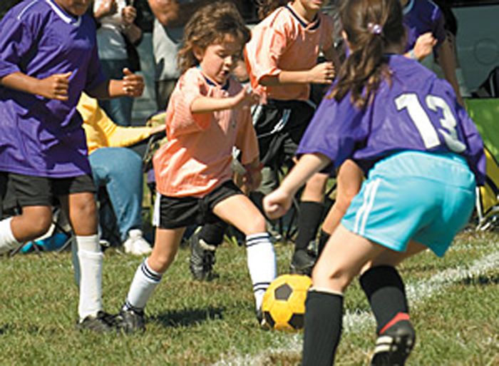
Physical activity is extremely important for everyone, but especially for children. A well-designed exercise program enhances the physical and intellectual development of a child. Competitive sports are often a child’s first introduction to programmed exercise.

In the past decade, there has been an increase in the number of children participating in team and solo sports. Younger children are allowed to participate in sports for enjoyment, health and personal development. However, this changes as competitive elements become more dominant and young athletes train harder and longer, and may practice a sport throughout the whole year. Consequently, sports-related injuries in children have significantly increased.

To understand pediatric injuries that can occur during sports performance, it’s important to be aware of the peculiarities of the growing musculoskeletal system. Children’s tendons and ligaments are relatively stronger than the epiphyseal plate; therefore, with severe trauma the epiphyseal plate will give way before the ligament. However, children’s bones and muscles are more elastic and heal faster. At the peak period of adolescent linear growth, the musculoskeletal system is most vulnerable because of imbalances in strength and flexibility and changes in the biomechanical properties of bone.

Physiological loading is beneficial for bones, but excessive strains may produce serious injuries to joints. Low-intensity training can stimulate bone growth, but high-intensity training can inhibit it. Growth plate disturbances resulting from sports injuries can result in limb-length discrepancy, angular deformity or altered joint mechanics, possibly causing permanent disabilities. Sports involving contact and jumping have the highest injury levels.

Pediatric Musculoskeletal Growth

Chiropractors have been uniquely trained to understand the musculoskeletal system, making them excellent resources for the management of sports-injuries. We need only to make certain we are aware of the peculiarities of the pediatric musculoskeletal system when pursuing appropriate evaluation and case management. (more…)

Introduction to the Dynamic Chiropractic Paradigm

By |June 14, 2011|Diagnosis, Education|

Introduction to the Dynamic Chiropractic Paradigm

The Chiro.Org Blog


We would all like to thank Dr. Richard C. Schafer, DC, PhD, FICC for his lifetime commitment to the profession. In the future we will continue to add materials from RC’s copyrighted books for your use.

The following is Chapter 1 from RC’s best-selling:

Motion Palpation

These materials are provided as a service to our profession. There is no charge for individuals to copy and file these materials. However, they cannot be sold or used in any group or commercial venture without written permission from ACAPress.


Chapter 1:   INTRODUCTION TO THE DYNAMIC CHIROPRACTIC PARADIGM

Overview of the Dynamic Chiropractic Approach

This chapter presents an overview of the background and basic concepts of Dynamic Chiropractic. The normal motions of spinal and related articulations, general considerations of spinal fixations, the different types of fixations, the significant physiologic mechanisms associated, a comparison of traditional and modern definitions of the vertebral subluxation complex, and other basic concepts are summarized.

In 1936, a small group of Belgium chiropractors began what was to be a long research project. Its aim was to study what chiropractors refer to as a subluxation, which is traditionally defined as an incomplete dislocation, a displacement in which the articular surfaces have not lost contact, or a partially reduced (spontaneously) dislocation.

Outstanding within the Belgium group were Drs. H. Gillet and M. Liekens. These investigators, who have been involved in this study for more than half a century, soon found that the clinical phenomenon of subluxation was a great deal more complicated than the effects of the oversimplified picture of “a bone out of place” that has been commonly proposed since the turn of the century. Their findings reported in the Belgium Research Notes are a testimony to their skillful observations. Although the theory of “a displaced vertebra” contained enough truth within it to constitute a basic therapeutic approach that could be justified by large numbers of positive benefits witnessed empirically, it was not sufficient to serve as a scientific hypothesis. (more…)

Alteration of Motion Segment Integrity

By |November 20, 2010|Diagnosis, Documentation, Education, Subluxation|

Alteration of Motion Segment Integrity

The Chiro.Org Blog


SOURCE:   Dynamic Chiropractic

By Jeffrey Cronk, DC, CICE


Sometimes the internal discourse that is common in our profession seems to get in the way of our acceptance of real help so that we can expand our profession and better serve our patients. Alteration of motion segment integrity (AOMSI) is a significant gift from the AMA that allows us to methodically locate, substantiate and objectively prove the severity of the spinal subluxation. Of course, it comes as a gift only as long as we handle it with a high level of responsibility.

Alteration of motion segment integrity is determined by exact mensuration procedure published in the AMA Guides to the Evaluation of Permanent Impairment. It is a spinal subluxation that can be objectively identified with a high degree of accuracy, especially when one acknowledges the advancements that have occurred in assessment of stress imaging (X-ray, DMX).

Please remember that some of the most significant advancements in functional radiology assessment came from information gained from our profession’s very first federal research grant, awarded in the mid 1970s. It was University of Colorado scientist Chung Ha Suh, PhD, who secured the first chiropractic funding from the National Institutes of Health (NS 12226 01A1). Suh’s main areas of research focused on the development of computerized, kinematic models of the spine and three-dimensional, distortion-free X-ray analysis. This research improved our ability to more accurately measure articular deformations such as AOMSI.

You may also want to review:

Accurate Prognosis in Personal-Injury Cases Using George’s Line

(more…)