Sports Management: Neck and Cervical Spine Injuries
We would all like to thank Dr. Richard C. Schafer, DC, PhD, FICC for his lifetime commitment to the profession. In the future we will continue to add materials from RC’s copyrighted books for your use.
This is Chapter 22 from RC’s best-selling book:
“Chiropractic Management of Sports and Recreational Injuries”
Second Edition ~ Wiliams & Wilkins
These materials are provided as a service to our profession. There is no charge for individuals to copy and file these materials. However, they cannot be sold or used in any group or commercial venture without written permission from ACAPress.
Chapter 22: NECK AND CERVICAL SPINE INJURIES
Soft-Tissue Injuries of the Posterior Neck
Cervical Contusions, Strains, and Sprains
Contusions in the neck are similar to those of other areas. They often occur to the cervical muscles or spinous processes. Painful bruising and tender swelling will be found without difficulty, especially if the neck is flexed. Phillips points out the necessity of normally lax ligaments at the atlanto-axial joints to allow for normal articular glidding, thus making tonic muscle action the only means by which head stability is obtained.
Strains (Grades 1–3) or indirect muscle injuries are common, frequently involving the erectors. Flexion and extension cervical sprains are also common in sports (Grades 1–3), and usually involve the anterior or posterior longitudinal ligaments, but the capsular ligaments may be involved. In the neck especially, strain and sprain may coexist. Severity varies considerably from mild to dangerous. Anterior injuries are more common to the head and chest as they project further anteriorly, but a blunt blow from the front to the head or chest may result in an indirect extension or flexion injury of the cervical spine. Many cervical strains heal spontaneously but may leave a degree of fibrous thickening or trigger points within the injured muscle tissue. Residual joint restriction following acute care is more common in traditional medical care than under mobilizing chiropractic supervision.
Cervical sprain and disc rupture are associated with severe pain and muscle spasm and are more common in adults because of the reduced elasticity of supporting tissues. Pain is often referred when the brachial plexus is involved. Cervical stiffness, muscle spasm, spinous process tenderness, and restricted motion are common. When pain is present, it is often poorly localized and referred to the occiput, shoulder, between the scapulae, arm or forearm (lower cervical lesion), and may be accompanied by paresthesias. Radicular symptoms are rarely present unless a herniation is present.
Diagnosis and treatment are similar to that of any muscle strain-sprain, but concern must be given to induced subluxations during the initial overstress. Palpation will reveal tenderness and spasm of specific muscles. In acute scalene strain, tenderness and swelling will usually be found. When the longissimus capitis or the trapezius are strained, they stand out like stiff bands.
Extension Injuries. When the head is violently thrown backwards (eg, whiplash), the damage may vary from minor to severe tearing of the anterior and posterior ligaments. Severe cord damage can occur which is usually attributed to momentary pressure from the ligamentum flavum and lamina posteriorly, even without roentgenographic evidence. A facial injury usually suggests an accompanying extension injury of the cervical spine as the head is forced backward. Management of minor injuries requires reduction of subluxations, traction, physiotherapeutic remedial aid, a supporting collar for as long as postural muscles are inadequate for structural support, followed by graduated therapeutic exercises.
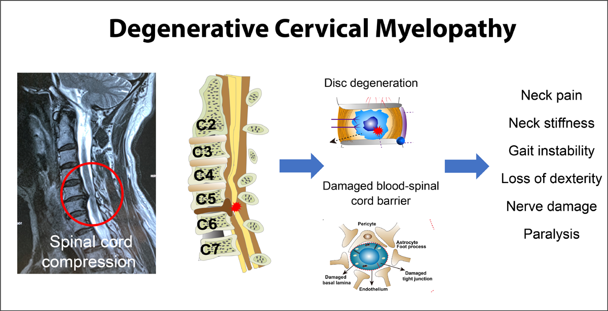
Flexion Injuries. Slight anterior subluxation is usually not serious, but neurologic symptoms may appear locally or down the arm. Disc degeneration may follow, leading to spondylosis. An occipital injury usually suggests an accompanying flexion injury of the cervical spine as the skull is forced forward. Management is similar to that of extension injuries except required support is often shorter (6–8 weeks).
Torticollis, Spasms, and Similar Disorders. Wry-neck spasm (tonic, rarely clonic) of the sternocleidomastoideus and trapezius may be due to irritation of the spinal accessory nerve by swollen glands, abscess, acute upper respiratory infections, scar, or tumor, but it more often occurs from traumatic cervical subluxations or idiopathically in “rheumatic” or “nervous” individuals. The muscles are rigid and tender, the head tilts toward the spastic sternocleidomastoideus, and the chin is rotated to the contralateral side. Common trigger points involved in “stiff neck” are in the trapezius (usually a few inches lateral to C7) or the levator scapulae and splenius cervicus lateral to C4–C6 cervical processes. These points are often not found unless the muscle is relaxed during palpation.
Wry neck may also be the result of subdiaphragmatic irritation being mediated reflexly into the trapezius and cervical muscles. Subclinical visceral irritation is often the factor involved.
Dislocations of upper cervical vertebrae cause a distortion of the neck much like that of torticollis. A fracture-dislocation of a cervical vertebra will produce neck rigidity and a fast pulse, but fever is absent. Local and remote trigger points are frequently involved. In suspicious cases, the neck should always be x-rayed before it is examined. Neck rigidity may also be the result of a sterile meningitis from blood in the cerebrospinal fluid. Thus, if a paatient has slight fever, rapid pulse, and rigid neck muscles, subarachnoid hemorrhage is suspected. Lateralizing signs are often indefinite.
Management. The benefits of rotary cervical adjustments are well known within the profession. To relieve muscle spasm, heat is helpful, but cold and vapocoolant sprays have shown to be more effective in acute cases. Mild passive stretch is an excellent method of reducing spasm in the long muscles. Heavy passive stretch, however, destroys the beneficial reflexes. For example, place the patient prone on an adjusting table in which the head piece has been slightly lowered. Turn the patient’s head toward the side of the spastic muscle. With head weight alone serving as the stretching force, the spasm should relax within 2–3 min. Thumb pressure, placed on a trigger area, is then directed towards the muscle’s attachment and held for a few moments until relaxation is complete.
Isotonic exercises are useful in improving circulation and inducing the stretch reflex, especially in the cervical extensors. These exercises should be done supine to reduce exteroceptive influences on the central nervous system.
Peripheral inhibitory afferent impulses can be generated to partially close the presynaptic gate by acupressure, acu-aids, acupuncture, or transcutaneous nerve stimulation. Most authorities feel deep sustained manual pressure on trigger points is the best method, but a few others prefer brutal short-duration pressure (1–2 sec). Deep pressure is contraindicated in any patient receiving anti-inflammatory drugs (eg, cortisone) as subcutaneous hemorrhage may result. The effects of cervical traction are often dramatic but sometimes short lived if a herniated disc is involved. In chronic cases, relaxation training with biofeeback is helpful.
An acid-base imbalance from muscle hypoxia and acidosis is frequently the cause which may be prevented by Lindahl’s alkalinization mixture (potassium citrate, 33.5%; calcium lactate, 41%; sodium citrate, 12%; magnesium glyconate, 12%; lithium citrate, 1.5%).
| Review the complete Chapter (including sketches and Tables) at the ACAPress website |


Leave A Comment