MRI Update
SOURCE: ACA News
By Caitlin Lukacs
Technological progress is not reserved for cell phones and iPads. Advanced imaging modalities, such as magnetic resonance imaging (MRI), have benefited from recent advances as well. In fact, MRI has become the gold standard of advanced imaging for the spine and extremity joints. “It’s hard to do anything in medicine without imaging,” says Norman Kettner, DC, DACBR, professor of clinical science and chairman of the Department of Radiology at Logan College of Chiropractic. “In fact, in a survey of the top 25 internists in the United States, MRI and CT scans were rated No. 1 as the most important development in medical science in the previous century.
What Is MRI?
Gary Longmuir, DC, DACBR, clinical radiologist and president of the ACA Council on Diagnostic Imaging, explains that MRIs create images of the internal structures of the body based on the energy released from hydrogen protons. Because the body is largely made up of water and each water molecule contains two hydrogen protons, when a person enters the magnetic field created by the MRI scanner, the magnetic charges of the protons change and align with the direction of the scanner’s field. When the field is then turned off, the protons return to their original energy state, releasing the energy difference as photons that are detected by the scanner as a “signal” similar to a radio wave. The protons of distinct tissues return to their original energy states at different rates, and this difference is detected by the MRI scanner.
DCs and MRI
“MRI is part of clinical evaluation,” explains Michael Fergus, DC, DACBR, who serves on the American Chiropractic College of Radiology and works as an instructor with the National University of Health Sciences. “It’s used to help diagnose injuries and illness based on what Dcs have found during the history and physical exam,” he continues. MRI can be used to image almost any part of the body – the central nervous system, the mediastinum/heart, parts of the abdomen, the biliary tract through the use of magnetic resonance cholangiopancreatography (MRCP), blood vessels through the use of magnetic resonance angiography (MRA), reproductive organs, spine and extremities.
“MRI is not meant as a scout study, though,” says Dr. Longmuir. “It’s either confirmatory or exclusionary. For example, an MRI can confirm a suspected cartilage tear in the knee or exclude a disc lesion.”
While there are no utilization guidelines specifically for doctors of chiropractic, the American College of Radiology (ACR) has created appropriateness criteria for MRIs that all practitioners should be familiar with, says Dr. Kettner. “For example, a patient comes in with chronic neck pain who has normal X-rays. You do a physical examination and find neurological symptoms. According to the ACR, that indicates a strong recommendation for MRI.”
Another example of when an MRI is strongly recommended is with a patient who has frequent headaches. If there is a change in the intensity of the headaches, or if another symptom suddenly appears, such as blurred vision, that would be an indication for MRI.
The appropriateness criteria can be found online at: www.acr.org
However, cautions Dr. Fergus, each case is unique, and the decision to use imaging should be made on a case-by-case basis. “There is no cookiecutter way to say every time you see this specific symptom, you should order an MRI,” he says.
Ordering Images
Once a DC has decided that an MRI is necessary, an image must be ordered from an imaging center or a hospital. Imaging centers are independent of hospitals and simply take the images and then send the results to a consultant to read. “A lot of conditions demand immediate attention on the part of the clinicians: brain bleeds, tumors, infective processes. For this reason, imaging consultants need to get the reports to the ordering doctors within 24 hours,” explains Dr. Longmuir, who works as a clinical radiologist for local imaging centers.
It’s not hard to find imaging centers to work with, says Dr. Kettner. “They’re always knocking on our door looking for a referral source because they want that patient volume,” he says. “But they have to do good scans and provide a safe and comfortable environment, and they’re usually very good about providing a good experience for the patient.” MRI center marketers approach Dcs-we don’t generally have to look for them, says Dr. Fergus. “They will bring their sales pitch and the appropriate paperwork to new doctors in the area.”
“The use of imaging equipment is a large motivation for Dcs to start making inroads into hospital systems, starting with outpatient privileging. Outpatient privileging would mean that the DC calls and puts the patient on the imaging schedule, the patient reports for the scan, the test is done and the interpretation is sent to the ordering doctor,” explains Dr. Kettner.
Imaging Studies
Most chiropractic colleges teach students enough about imaging to allow them to discuss reports with their patients. DCs are trained to understand the report and how it will affect the treatment of their patients, explains Dr. Kettner. They should also be able to identify a disc protrusion or stenosis, for example, he says. However, Dcs shouldn’t stray too far from a consulting radiologist, Dr. Kettner cautions.
At National, we have a course that runs through advanced orthopedic imaging,” explains Dr. Fergus. “Basically we cover various orthopedic conditions and how they would appear in advanced imaging, as well as which imaging modalities are appropriate to order in each situation.”
For those DCs who have an interest in imaging and want to further their education, a diplomate program is available. “Residents in radiology study imaging in every system of the body, says Dr. Kettner. “Radiology residency training is magnified 100 times over the training in the doctoral program itself,” he explains.
Becoming a diplomate in diagnostic imaging opens up additional professional opportunities, says Dr. Fergus. “In addition to working in private practice, a DC could pursue a career in education, work as a consultant with an imaging center and network with other types of physicians,” he says.
The Future of Imaging
Until now, imaging has been limited to the anatomical level of the body. The future of imaging is aimed at defining how things function at the molecular level, says Dr. Longmuir. “The potential uses for this type of imaging are numerous. Molecular imaging could be used to detect cancer markers in proteins of specific cells.”
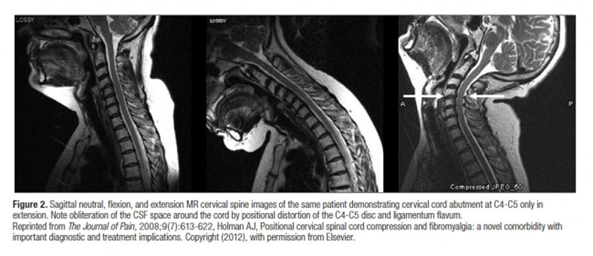
Another new type of imaging is weight-bearing MRIs. Fonar Corp. has been conducting studies in MRIs in the weight-bearing seated and standing postures. “Weight-bearing imaging makes certain functional pathologies more obvious. For example, cervical images in full flexion and full extension can show anterior and posterior spinal displacement or disc bulges and protrusions that are not present in MRIs taken in the recumbent position,” says Dr. Longmuir.


Leave A Comment