
Real-Time Visualization of Joint Cavitation
SOURCE: PLoS One. 2015 (Apr 15); 10 (4): e0119470
Gregory N. Kawchuk, Jerome Fryer, Jacob L. Jaremko,
Hongbo Zeng, Lindsay Rowe, Richard Thompson
Department of Physical Therapy,
Faculty of Rehabilitation Medicine,
University of Alberta,
Edmonton, Alberta, Canada
Cracking sounds emitted from human synovial joints have been attributed historically to the sudden collapse of a cavitation bubble formed as articular surfaces are separated. Unfortunately, bubble collapse as the source of joint cracking is inconsistent with many physical phenomena that define the joint cracking phenomenon. Here we present direct evidence from real-time magnetic resonance imaging that the mechanism of joint cracking is related to cavity formation rather than bubble collapse. In this study, ten metacarpophalangeal joints were studied by inserting the finger of interest into a flexible tube tightened around a length of cable used to provide long-axis traction. Before and after traction, static 3D T1-weighted magnetic resonance images were acquired. During traction, rapid cine magnetic resonance images were obtained from the joint midline at a rate of 3.2 frames per second until the cracking event occurred. As traction forces increased, real-time cine magnetic resonance imaging demonstrated rapid cavity inception at the time of joint separation and sound production after which the resulting cavity remained visible. Our results offer direct experimental evidence that joint cracking is associated with cavity inception rather than collapse of a pre-existing bubble. These observations are consistent with tribonucleation, a known process where opposing surfaces resist separation until a critical point where they then separate rapidly creating sustained gas cavities. Observed previously in vitro, this is the first in-vivo macroscopic demonstration of tribonucleation and as such, provides a new theoretical framework to investigate health outcomes associated with joint cracking.
Enjoy this live video demonstration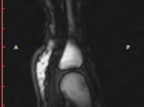 |
From the FULL TEXT Article:
Introduction
Background
Sounds emitted from human synovial joints vary in their origin. Joint sounds that occur repeatedly with ongoing joint motion arise typically when anatomic structures rub past one another. In contrast, “cracking” sounds require time to pass before they can be repeated despite ongoing joint motion. Although various hypotheses have been proposed over many decades regarding the origin of cracking sounds, none have been validated; the underlying mechanism of cracking sounds remains unknown.
History
In 1947, Roston and Wheeler Haines [1] published the first scientific study toward describing the origins of joint cracking. Their experiment used serial radiography to visualize joint cracking when distraction forces were applied to metacarpophalangeal (MCP) joints. Their results characterized the sequence of gross articular events that define joint cracking. The process begins with the resting phase where joint surfaces are in close contact. In this stage, a light distraction force will barely separate the joint surfaces. With a greater distraction force, the surfaces resist separation until a critical point after which they separate rapidly. It is during this rapid separation phase that the characteristic cracking sound is produced. Following cracking, the joint is in a refractory phase where no further cracking can occur until time has passed (approximately 20 minutes). Importantly, post-cracking distraction also reveals the presence of a “clear space” assumed by Roston and Wheeler Haines to be a vapour cavity. This cavity, described by some as a bubble, has been thought to form as distraction forces decrease pressure within the synovial fluid to the point were dissolved gas comes out of solution. Importantly, Roston and Wheeler Haines linked the production of the cracking sound to the formation of this clear space, a phenomenon first described in 1911 [2] but thought by some to occur only in unhealthy joints [3] until demonstrated to also occur in normal joints [4].
Read the rest of this Full Text article now!


Here is a gif of the MRI…
I’ve posted an animated gif of the cavitation in the comments.
Thanks John!
Great realisation ! Would like to show it on my next healthtalk…h