
Patient with Low Back Pain and Somatic Referred Pain Concomitant with Intermittent Claudication in a Chiropractic Practice
SOURCE: Topics in Integrative Health Care 2014 (Mar 27); 5 (1)
Kathryn Hoiriis, D.C., Brent S. Russell, M.S., D.C.
Introduction: Approximately 12% of older patients in the general population have atherosclerotic disease of the aorta and lower extremity arteries, i.e., peripheral artery disease (PAD). Intermittent claudication is the most common symptom. When a patient with low back pain complains of lower extremity pain that is worsened with mild exercise (e.g. walking), the etiology is often not clear.
Case Presentation: A 56 year-old male presented with low back pain, left hip and buttock discomfort, numbness in thigh and calf, and left knee weakness while walking.
Intervention and Outcome: Chiropractic care was provided and the low back pain improved. The patient developed leg weakness. Radiographic evaluation showed calcification of abdominal aorta and common iliac arteries. The patient was referred for medical evaluation and diagnostic ultrasound findings of arterial occlusion lead to surgical referral. The surgeon reported a “significant amount” of blockage of the left external iliac artery. Leg weakness resolved following placement of surgical stents.
Discussion: Claudication may go undiagnosed because many people consider the pain a consequence of aging, and may therefore just reduce their activity level to avoid the pain. Early diagnosis of PAD/intermittent claudication is important since PAD is a major risk factor for adverse cardiovascular events.
There are more articles like this @ our:
Conclusion: Patient management in the chiropractic clinical setting required appropriate medical referral in this case. Surgical implantation of stents in the left external iliac artery resolved the complaint of leg weakness. It is imperative for health care professionals to have awareness of the high occurrence of PAD in the general population.
From the FULL TEXT Article
Introduction
Claudication is defined as pain caused by too little blood flow within muscles of the lower extremity. The pain is often described as intermittent or “on and off”. [1-3] Often, it is in the legs, but the arms can be affected. [4] The pain may come on during mild exercise, such as walking a specified distance, and typically subsides with rest. Claudication is a symptom of a disease, peripheral artery disease (PAD), which is potentially serious circulation problem. Comerata defines PAD as atherosclerotic disease of the aorta and arteries of the lower extremities and states that the most frequent manifestations of ischemia occur in the lower extremity arteries, with intermittent claudication as the most common symptom. [3]
Approximately 12% of patients in the general population over the age of 50 have lower extremity PAD; [5] the incidence rises to about 20% by age 70, [6] and risks are also increased by smoking and diabetes. [5, 7-9] The diagnosis of PAD is considered to be a major risk factor for future cardiovascular events and increased mortality. [1, 10] The risk of mortality in patients with asymptomatic PAD is reported by investigators to be similar to those with severe or symptomatic PAD. [1, 2, 10-13] It has not been clearly determined how long the underlying pathophysiology may exist as asymptomatic PAD before becoming symptomatic with claudication. [11] The problem may gradually worsen until the pain occurs at rest. Signs of progression of the underlying pathology include discolored skin or ulcerations and, if blood flow is severely reduced, toes or fingers may look bluish and feel cold to the touch. Other possible symptoms include aching, burning, or weakness. Claudication and peripheral artery disease can reduce quality of life by limiting participation in social and leisure activities, interference with work, make exercise intolerable and may lead to potentially life-threatening complications if left untreated. The health-related quality of life of patients with PAD is reported to be similar to patients with other forms of cardiovascular disease, and PAD has been associated with high rates of depression. [5, 14, 15]
With the frequency of the problem of PAD in the population, it is important to raise awareness of health care professionals for appropriate differential diagnosis, clinical management and referral. Claudication isn’t the only possible cause of leg pain. Other conditions associated with similar symptoms that need to be considered include spinal stenosis, peripheral neuropathy, certain musculoskeletal conditions and deep venous thrombosis. This case report describes a 56 year old man with a history of low back injury, recurrent pain and leg pain.
Case Presentation
The patient is a 56 year-old male who presented for chiropractic care in December 2011, complaining of low back pain, discomfort in the left hip and buttock area, occasional numbness in his distal thigh and calf, and tingling/numbness in the distal anterior thigh, and left knee weakness while walking. These symptoms were not unusual or unexpected for this patient due to prior care for spinal complaints. A long case history of low back and leg injuries included hospitalization at age 21 with a disc herniation following a lifting injury, injections for low back pain at age 29, several occasions of pain due to yard work including an incident in which he fell backwards into a woodpile, and an episode of severe pain following a strained left hamstring muscle. He reported being a ‘light” cigarette smoker, “off and on” for many years, and also being diligent about his nutritional and exercise habits.
Initial Examination Findings: During the examination for this episode, Straight Leg Raise test caused thigh pain, although Bragard’s test was negative. The patient complained of general stiffness during lumbar range of motion. He was limited in lumbar extension, which also elicited a moderate pain response. Manual muscle testing revealed weakness (grade 4) of the left toe extensors, psoas, rectus femoris, and piriformis muscles. Other orthopedic and neurological tests yielded unremarkable (e.g. negative) findings.
Intervention and Outcomes
Chiropractic Care and Management: On the initial visit, he received lumbar and sacroiliac manipulations, along with flexion distraction. The patient did not return until 6 weeks later, at which time he reported improvement for his low back complaint. He reported continued weakness in his left knee, and occasionally felt tired during long walks. Resting helped so that he could continue walking. He noticed the same pattern while working in the yard, especially when he climbed up and down hills.
At the second chiropractic visit, examination included palpation for the posterior tibial, dorsal pedal, and popliteal pulses. These pulses were found present bilaterally; however, examination of the femoral pulses was not done. Foot skin color appeared equal left to right, with neither side appearing pale. Although there were clues that he might have vascular intermittent claudication, he also had neuromusculoskeletal signs and symptoms. The patient also reported he had described his symptoms to his internist, who examined him and did not think he had a serious problem; he had been instructed to return if the symptoms persisted or worsened.
The patient was seen for three additional chiropractic visits to manage neuromusculoskeletal dysfunction, and he received spinal manipulations, instruction on low back exercises, and transverse friction massage to muscles of the knee (popliteus, short head of the biceps femoris, gastrocnemius, rectus femoris.) He felt improved during this time, and he reported at the third visit he no longer needed to stop when he felt weakness, just to slow down. At the fourth visit, he complained of weakness in his left hip and buttock area, weakness and tiredness in his leg as before, and some burning in the bottom of his left foot. Upon examination, pulses were present bilaterally and foot coloration appeared bilaterally equal.
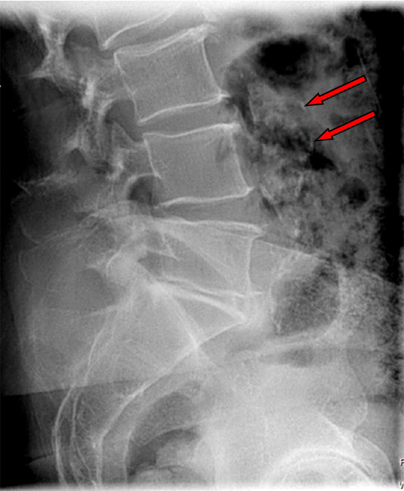
Radiographic Study: A clinical decision to obtain a lumbar radiographic study was made at the sixth visit and the patient was referred for digital radiographic imaging. Antero-Posterior (A-P), Lateral and L5 Spot radiographic images were obtained. These views demonstrated signs of degeneration at several levels of the lumbar spine including loss of disc heights, facet joint narrowing, bony proliferation, reactive sclerosis, and a degenerative retrolisthesis of L2. Significant calcification was noted within the abdominal aorta and common iliac arteries. (Figure 1)
Figure 1. Radiographic Image with Calcification of Abdominal Aorta
Legend: Red arrows indicate location observed calcification in Abdominal Aorta in the Lateral Lumbopelvic View.
Referral: The patient was instructed to return to his internist, and was sent for an ultrasound evaluation. The test showed signs of “hemodynamically significant occlusive disease” in the left leg, suggestive of “iliac or iliofemoral disease…in a range consistent with claudication”.
Surgical Intervention and Management: The patient had two stents inserted into the left external iliac/femoral artery, approximately four months after the office visit in which leg weakness was found. The surgeon reported a “significant amount” of blockage of the artery. After 6 weeks of surgical recovery, the leg weakness was resolved, though the patient complained of a discomfort in his groin from the stents. The patient began taking Plavix. He also began taking various nutritional supplements that he had read may help prevent future vascular problems, and he quit smoking cigarettes. At 20 months after the surgery, the patient reported that he feels fine. He no longer has the groin discomfort or any other complications. He has not resumed smoking.
Discussion
Case reports found in the literature described the clinical features of both arterial and neurogenic intermittent claudication. [16-20] Wiles and Blackmore have described issues with clinical diagnosis and management of claudication. [21] Claudication may go undiagnosed because many people consider the pain an unwelcome but inevitable consequence of aging, and some people just reduce their activity level to avoid the pain. Other conditions, such as spine, joint, or muscle problems can cause pain in legs. Diagnosis is based on symptoms and a medical history, physical exam and appropriate tests. The pain occurs intermittently because the leg muscles are not getting enough oxygenated blood to contract properly during exercise. PAD is usually the result of atherosclerosis. Atherosclerotic plaques cause hardening and narrowing of the arterial lumen thereby reducing the flow of blood. The risk factors for claudication are the same as those for developing atherosclerosis. (Figure 2) Smoking significantly increases the risk of PAD to 30%. [11, 12]
If your patient is…
|
If the patient’s systolic blood pressure at the ankle is less than 90% of the blood pressure in the arm, there’s a good chance of PAD or other atherosclerotic conditions, whether or not the patient is symptomatic.
|
Since peripheral arterial disease is a major risk factor for adverse cardiovascular events, Comerota and others advocate for active screening and early diagnosis of PAD/intermittent claudication. [3, 5, 11] Common tests that are widely used and are well described in the medical literature include pulses in the feet, ankle-brachial index (ABI), Doppler ultrasound, magnetic resonance imaging (MRI), computed tomography (CT) angiography. [5, 11, 21, 22] The literature provided a discussion of the usefulness of these tests. [3, 5, 11] Collins, et al, concluded that peripheral pulse palpation alone is not adequate as a screening tool for PAD, finding that more than 2/3 of a patient group with PAD had detectable pulses. [23]
The Ankle-Brachial Index has been established as an uncomplicated and inexpensive way to determine whether a patient is likely to have PAD. [23, 24] It is reliable when performed by trained and experienced personnel. [25, 26] It is highly specific (better than 90%) and has good sensitivity (less than 80%). [27] The ABI is a ratio of the systolic blood pressure taken from the ankle to the systolic pressure taken from the brachial artery. Pressures should be measured for both the posterior tibial and dorsalis pedis arteries, and use of a stethoscope is unacceptable; [27] a hand-held continuous wave Doppler ultrasound device should be used. [24, 27, 28] The ABI is calculated by using the highest systolic pressure of the 4 measured at the ankles, and divided by the higher systolic pressure for the left and right brachial arteries. [23, 24, 28] An ABI of less than 0.9 indicates the presence of PAD or other atherosclerotic conditions, whether or not the patient is symptomatic. [27, 29]
Treatment of claudication and peripheral artery disease can help prevent disease progression and reduce symptoms. Aggressive medical management incorporates risk factor modification and exercise therapy in addition to platelet inhibition and other pharmacotherapy. [3, 30, 31] Important in overall patient management are necessary lifestyle changes. Quitting smoking and participating in a regular exercise regimen are often the first steps in treating claudication. If claudication symptoms do not improve with these changes, then other treatment options include medications, angioplasty and vascular surgery.
The patient experienced a successful outcome with surgery in this case and also chose to quit smoking cigarettes. He has included various nutritional supplements. It is unclear as to which nutritional supplements are helpful. A systematic review of the literature reported that Ginkgo, Propionyl-L-carnitin, Vitamin E and omega-3 fatty acids as nutritional supplements did not have strong evidence of effectiveness for treatment of claudication. [32] Studies included in the review on the use of Ginkgo and Propionyl-L-carnitin had mixed results. The review suggested L-arginine may be helpful in relieving symptoms of claudication, but there were some questions about adverse reactions. [32] Vitamin E and omega-3 fatty acids have been suggested as treatments for claudication, but clinical trials that demonstrate significant beneficial changes in symptoms are needed. [32]
Conclusion
This case report illustrates that peripheral arterial disease can present with symptoms of backache and aching legs which are symptoms of intermittent claudication, and therefore, a thorough examination to make the appropriate clinical decision is critical. This report may raise the awareness of health care professionals about the high incidence of PAD in the population as well as provide some of the strengths and limitations of various screening methods for PAD.
References
- Alzamora MT, Baena-Díez JM, Sorribes M, Forés R, Toran P, Vicheto M, et al.
Peripheral Arterial Disease Study (PERART): Prevalence and predictive values of asymptomatic peripheral arterial occlusive disease related to cardiovascular morbidity and mortality for the PERART study.
BMC Public Health. 2007,7:348
available from http://www.biomedcentral.com/1471-2458/7/348 - Hirsch AT, Criqui MH, Treat-Jacobson D, Regensteiner JG, Creager MA, Olin JW, et al.
Peripheral arterial disease detection, awareness, and treatment in primary care.
JAMA. 2001;286:1317-24. - Comerota AJ.
The case for early detection and integrated intervention in patients with peripheral arterial disease and intermittent claudication.
J Endovasc Ther 2003;10:601-13.
Available from: www.jevt.org - Moritz G, Pollard H, Rigby S.
Subclavian Steal Syndrome: a review.
Chiropr Osteopat. 1998;7(1):20-8. - Armstrong DWJ, Tobin C, Matangi MF.
The accuracy of the physical examination for the detection of lower extremity peripheral arterial disease.
Can J Cardiol. 2010;26(10):e346-50. - Regensteiner JG, Hiatt WR.
Current medical therapies for patients with peripheral arterial disease: a critical review.
Am J Med. 2002;112(1):49-57. - Criqui MH, Langer RD, Fronek A, Feigelson HS, Klauber MR, McCann TJ, et al.
Mortality over a period of 10 years in patients with peripheral arterial disease.
N Engl J Med. 1992;326:381-6. - Fowkes FGR, Housely E, Cawood EHH, MacIntyre CCA, Ruckley CV, Prescott RJ.
Edinburgh Artery Study: Prevalence of asymptomatic and symptomatic peripheral arterial disease in the general population.
Int J Epidemiol. 1991;20:384-92. - Doobay AV, Anand SS.
Sensitivity and specificity of the ankle-brachial index to predict future cardiovascular outcomes: A systematic review.
Arterioscler Thromb Vasc Biol. 2005;25:1463-9.
Available from http://atvb.ahajournals.org/content/25/7/1463 - Leng GC, Lee AJ, Fowkes FG, Whiteman M, Dunbar J, Housley E, et al.
Incidence, natural history and cardiovascular events in symptomatic and asymptomatic peripheral arterial disease in the general population.
Int J Epidemiol. 1996;25:1172-81. - Mohler ER, Bundens W, Denenberg J, Medenilla E, Hiatt WR, Criqui MH.
Progression of asymptomatic peripheral artery disease over 1 year.
Vasc Med. 2012;17(1):10-6. - Ain DL, Slovut DP, Kamath R, Jaff MR.
The association between peripheral artery and lumbar spine disease: a single-center study.
Am J Med. 2012;125(4):411-5. - Diehm C, Allenberg JR, Pittrow D, Mahn M, Tepohl G, Haberl RL, et al.
Mortality and vascular morbidity in older adults with asymptomatic versus symptomatic peripheral artery disease.
Circulation. 2009;120:2053-61.
Available from: http://circ.ahajournals.org/content/120/21/2053 - Regensteiner JG, Hiatt WR, Coll JR, Criqui MH, Treat-Jacobson D, McDermott MM, et al.
The impact of peripheral arterial disease on health-related quality of life in the Peripheral Arterial Disease Awareness, Risk, and Treatment: New Resources for Survival (PARTNERS) Program.
Vasc Med. 2008;13:15-24. - McDermott MMG, Greenland P, Guralnik JM, Liu K, Criqui MH, Pearce WH, et al.
Depressive symptoms and lower extremity functioning in men and women with peripheral arterial disease.
J Gen Intern Med. 2003;18:461-7. - Henderson DJ.
Intermittent claudication – with special reference to its neurogenic form as a diagnostic and management challenge.
J Canadian Chiropr Assoc. 1979;23(1):9-19. - Thiel HW, Mior SA.
Coexistent vascular and spinal claudication: a report of two cases.
J Canadian Chiropr Assoc. 1987;31(3):131-6. - Downs SE.
Unilateral intermittent claudication of the left lower extremity.
J Manipulative Physiol Ther. 1988;11(4):322-4. - Sandoz R.
The narrow lumbar canal of degenerative origin. A case presentation.
Ann Swiss Chiropr Assoc. 1989;9:91-125. - Wessely MA, Garnesson C, Guenoun O:
Imaging case challenge.
J Acad Chiropr Orthoped. 2011;8(2):3-6. - Wiles M, Blackmore T.
Clinical diagnosis of peripheral vascular disease.
J Canadian Chiropr Assoc. 1978;22(3):101-5. - Terenzi T, Gallagher D, DeMeersman R, Beadle E, Muller D.
The age-related advancement of arterial disease measured by Doppler ultrasound diastolic flow analysis.
J Manipulative Physiol Ther. 1993;16(8):527-36. - Collins TC, Suarez-Almazor M, Peterson NJ.
An absent pulse is not sensitive for the early detection of peripheral arterial disease.
Fam Med. 2006;38(1):38-42. - Olin JW, Sealove BA.
Peripheral artery disease: current insight into the disease and its diagnosis and management.
Mayo Clin Proc. 2010;85(7):678-92. - Kaiser V, Kester AD, Stoffers HE, Kitslaar PJ, Knottnerus JA.
The influence of experience on the reproducibility of the ankle-brachial systolic pressure ratio in peripheral arterial occlusive disease.
Eur J Vasc Endovasc Surg. 1999;18(1):25-9. - Mätzke S, Franckena M, Albäck A, Railo M, Lepäntalo M.
Ankle brachial index measurements in critical leg ischaemia – the influence of experience on reproducibility.
Scand J Surg 2003;92(2):144-7. - Aboyans V, Criqui MH, Abraham P, Allison MA, Creager MA, Diehm C, et al.
Measurement and interpretation of the Ankle-Brachial Index: a scientific statement from the American Heart Association.
Circulation. 2012;126:2890-909. - Nicolaï SPA, Kruidinier LM, Rouwet EV, Bartelink MLEL, Prins MH, Teijink JAW.
Ankle brachial index measurement in primary care: are we doing it right?
Br J Gen Pract. 2009;59:422-7. - Fowkes FG, Murray GD, Butcher I, Heald CL, Lee RJ, Chambless LE, et al.
Ankle brachial index combined with Framingham Risk Score to predict cardiovascular events and mortality: a meta-analysis.
JAMA. 2008;300(2):197-208. - Burns P, Lima E, Bradbury AW.
What Constitutes Best Medical Therapy for Peripheral Arterial Disease?
Eur J Vasc Endovasc Surg. 2002;24(6):12 pages.
Available from: http://www.idealibrary.com - DeVries SO, Visser K, deVries JA, Wong JB, Donaldson MC, Hunink, MGM
Intermittent claudication: cost-effectiveness of revascularization versus exercise therapy.
Radiology. 2002;222:25-36.32. Pittler MH, Ernst E.
Complementary therapies for peripheral arterial disease: systematic review.
Atherosclerosis. 2005;181(1):1-7.
Available from: http://www.atherosclerosis-journal.com
Leave A Comment